Abdominal ultrasound is helpful in diagnosing midgut malrotation and volvulus in pediatric patients, according to research presented November 30 at the RSNA annual meeting.
Dr. Haithuy Nguyen from Texas Children's Hospital presented findings from her team's multicenter study of over 550 pediatric patients that showed high sensitivity and specificity in imaging these abdominal conditions.
"Abdominal ultrasound is increasingly being clinically used to evaluate for midgut malrotation and volvulus," Nguyen said.
Malrotation occurs when the intestine does not turn like it should, which can happen early in pregnancy. This affects about one out of every 500 children in the U.S. This can result in volvulus, in which the intestine becomes twisted, causing blockage.
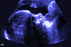
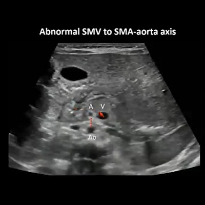
Midgut malrotation (above) and volvulus (below) can be found in pediatric patients with abdominal abnormalities. Ultrasound shows high sensitivity and specificity in diagnosing these conditions, according to research led by Dr. Haithuy Nguyen from Texas Children's Hospital. Images courtesy of Dr. Haithuy NguyenUltrasound has been touted by researchers as being noninvasive and emitting no radiation, making it suitable for young children.
Nguyen et al wanted to report the sensitivity and specificity of ultrasound for both abdominal conditions separately and in the clinical setting.
They used retrospective data from three tertiary children's hospitals and included 551 children in the study ranging from ages 0 to 18 years. They were evaluated by ultrasound between 2018 and 2021. Children with previously known malrotation or normal rotation were excluded.
Out of the 551 children included, 283 were male and 268 were female, and the median age range was 1.5 months. A total of 574 ultrasound exams were collected.
Ultrasound showed a sensitivity of 93% and a specificity of 95% when it came to imaging diagnostic malrotation. For equivocal tests, ultrasound's sensitivity did not increase while specificity dropped to 87%.
"If you took all of those [equivocal tests] and assumed them to be negative, then it dropped the sensitivity to 89% and increased specificity only marginally to 96%," Nguyen said.
For volvulus, ultrasound had perfect sensitivity (100%) and 98% specificity on diagnostic tests. For equivocal tests, ultrasound retained perfect sensitivity, but it dropped to 95% specificity. If all equivocal tests were assumed to be negative, sensitivity dropped to 85% while specificity increased to 98%.
Nguyen said while about one-third of the tests included were nondiagnostic or did not properly report malrotation or volvulus, one-fourth of those did report alternative findings.
"Additional findings and alternative diagnoses are not uncommon, so ultrasound does provide that additional angle," she added.
Nguyen also said the team looked at patient outcomes after imaging and is submitting another abstract related to that. She said the speed of imaging and admittance to the operating room did not achieve statistical significance, but the researchers noticed a trend where admittance to the operating room was faster.
"Even if we had to do an upper GI, a lot of the information was already there, but we just needed to prove to the surgeon that there was malrotation," she said. "The radiologist already knew the answer and the performing surgeon had a feeling it was the answer. So, everything just sped up regardless."
Nguyen also said another trend noticed was the length of hospital stay decreased and less complications were seen. However, these data points did not achieve statistical significance. Nguyen said this may be due to the small sample size.



![Examples of ultrasound findings and techniques. (A) Images in a 39-year-old male patient with a mass in the left thigh. The mass is heterogeneous on the B-mode US image (compared with the patient in D) and showed increased microvascularity (superb microvascular imaging [SMI]) and shear-wave elastography (SWE) values. Undifferentiated pleomorphic sarcoma was diagnosed at biopsy (with pleomorphic rhabdomyosarcoma in surgical specimen). (B) Images in an 18-year-old male patient with a mass in the left leg. The mass is hypoechoic on the B-mode image, with no other findings suggestive of malignancy. The lesion is in contact with the cortex of the tibia, which is slightly irregular. CT revealed a doubtful anteromedial tibial erosion. The microvascular study demonstrated high vascularization, suggestive of malignancy. Periosteal Ewing sarcoma was diagnosed with both histologic and immunohistochemical confirmation. (C) Images in a 69-year-old female patient with a lump growing on the outside of the left leg. Multiple SWE examinations were performed (please note the high values obtained in the measurements, whereas the color map highlights the stiffness relative to adjacent tissues). SMI showed areas of increased vascularization to target for sampling. Undifferentiated spindle cell sarcoma was diagnosed at biopsy, with residual leiomyosarcoma in the surgical specimen after neoadjuvant therapy. (D) Images in a 56-year-old female patient with a mass in the right thigh. The mass is heterogeneous at both B-mode ultrasound (similar to patient A) and MRI (coronal T2-weighted spectral attenuated inversion recovery [SPAIR]; T1-weighted pre-contrast and postcontrast imaging), which even shows uptake after the administration of paramagnetic contrast material, which is traditionally suggestive of malignancy. Low values at SMI and elastography are suggestive of benignity. Spindle cell lipoma was diagnosed at biopsy, with atypical spindle cell lipomatous tumor in the surgical specimen.](https://img.auntminnie.com/mindful/smg/workspaces/default/uploads/2026/08/images-radiol250278fig2.APCFLSvX6p.jpg?auto=format%2Ccompress&fit=crop&h=100&q=70&w=100)


![Examples of ultrasound findings and techniques. (A) Images in a 39-year-old male patient with a mass in the left thigh. The mass is heterogeneous on the B-mode US image (compared with the patient in D) and showed increased microvascularity (superb microvascular imaging [SMI]) and shear-wave elastography (SWE) values. Undifferentiated pleomorphic sarcoma was diagnosed at biopsy (with pleomorphic rhabdomyosarcoma in surgical specimen). (B) Images in an 18-year-old male patient with a mass in the left leg. The mass is hypoechoic on the B-mode image, with no other findings suggestive of malignancy. The lesion is in contact with the cortex of the tibia, which is slightly irregular. CT revealed a doubtful anteromedial tibial erosion. The microvascular study demonstrated high vascularization, suggestive of malignancy. Periosteal Ewing sarcoma was diagnosed with both histologic and immunohistochemical confirmation. (C) Images in a 69-year-old female patient with a lump growing on the outside of the left leg. Multiple SWE examinations were performed (please note the high values obtained in the measurements, whereas the color map highlights the stiffness relative to adjacent tissues). SMI showed areas of increased vascularization to target for sampling. Undifferentiated spindle cell sarcoma was diagnosed at biopsy, with residual leiomyosarcoma in the surgical specimen after neoadjuvant therapy. (D) Images in a 56-year-old female patient with a mass in the right thigh. The mass is heterogeneous at both B-mode ultrasound (similar to patient A) and MRI (coronal T2-weighted spectral attenuated inversion recovery [SPAIR]; T1-weighted pre-contrast and postcontrast imaging), which even shows uptake after the administration of paramagnetic contrast material, which is traditionally suggestive of malignancy. Low values at SMI and elastography are suggestive of benignity. Spindle cell lipoma was diagnosed at biopsy, with atypical spindle cell lipomatous tumor in the surgical specimen.](https://img.auntminnie.com/mindful/smg/workspaces/default/uploads/2026/08/images-radiol250278fig2.APCFLSvX6p.jpg?auto=format%2Ccompress&fit=crop&h=112&q=70&w=112)