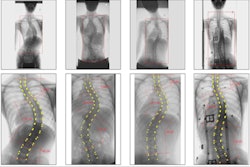
Ultrasound is useful in identifying schoolchildren who do not require specialist referral for follow-up x-rays as part of scoliosis screening programs, according to research published June 28 in Ultrasound in Medicine and Biology.
Researchers led by Dr. Henry Pang from the Chinese University of Hong Kong found high accuracy for ultrasound in determining the threshold of referral that would require x-ray for children with signs of scoliosis. Using ultrasound could reduce unnecessary x-ray exposure for children participating in scoliosis screening programs.
"Ultrasound could be considered for incorporation into the scoliosis screening program for maintaining radiation exposure as low as reasonably achievable, especially for immature participants," the study authors wrote.
Screening children for scoliosis is important for timely brace treatment to lessen spinal curvature. In Hong Kong, children who screen positive with a scoliometer and Moire topography (which presents a 3D representation of the back) are typically referred to x-ray.
Despite x-ray's high sensitivity in detecting children who need specialist referral, radiography also has a false-positive rate of more than 50%, which could lead to unnecessary radiation exposure.
On the other hand, ultrasound does not expose patients to radiation, and it has shown promise in assessing how severe spinal curvature is in scoliosis patients. Therefore, the researchers wanted to see how accurate ultrasound was in determining the threshold of referral that requires x-ray for children who test positive at screening.
The study included 442 schoolchildren in Hong Kong with an average age of 13.2 years. They had an average side-to-side spinal angle (Cobb angle) of 14.0° ± 6.6°.
In predicting the correct referral status for patients, ultrasound showed a sensitivity of 92.3% and a specificity of 51.6%, with positive and negative predictive values of 29% and 96.9%, respectively. The results were confirmed by x-ray.
The area under the curve (AUC) value for ultrasound alone was 0.735, while combining ultrasound with measuring angle of trunk rotation boosted the AUC to 0.832.
"In other words, if incorporated as an integral part of the scoliosis screening program, when schoolchildren are screened positive but before confirmative radiographic examination, ultrasound can prevent unnecessary exposure to radiation from confirmatory radiography in around 50% of cases for those with radiological Cobb angles below the specialist referral threshold of 20°," the researchers wrote.
The team also suggested that along with referral status, ultrasound in scoliosis could be extended by establishing a model for predicting the Cobb angle based on the angle of spinous processes, bony projections on the back of each vertebra. However, more studies would be needed to confirm this.
The authors also stated that improving ultrasound scanners to more accurately examine more spinal curves and expand to more regions of the back, such as the lower cervical region and sacral area, would reduce false-negative outcomes.