
Orthopedic surgeons in Japan have developed an AI method based on spine x-rays that could be used to help evaluate scoliosis, according to a study published September 4 in Scientific Reports.
A team led by Yoshihiro Maeda, PhD, of Keio University in Tokyo, trained a deep-learning algorithm to detect vertebrae and automatically measure spine curves on x-ray images of patients with adolescent idiopathic scoliosis (AIS). In testing, the method compared well with evaluations by expert doctors, they found.
"The proposed method showed a high correlation with the doctors' measurements, regardless of the [Cobb angle] size, doctors' experience, and patient posture," the group wrote.
Children between 10 and 17 years old who have scoliosis of unknown cause are classified as having AIS, with standing whole-spine x-ray as the standard diagnostic imaging technique. Surgery is typically based on the severity of the spinal deformity, which is indicated by the Cobb angle, a measurement between the two lines of the vertebral endplates at the upper and lower ends of the curve.
Years ago, doctors measured Cobb angles to diagnose scoliosis using a protractor on printed x-ray images, with scoliosis considered mild with a Cobb angle less than 20° and severe with Cobb angles greater than 50°. Most PACS now have built-in features that automatically calculate the angle, yet PACS still require manual selection of the appropriate end vertebrae by surgeons, the researchers noted.
As a potential aid in these cases, the researchers aimed to train and evaluate a convolutional neural network (CNN) that can automatically measure the Cobb angle.
In training, they used 1,021 full-length x-ray images of the spines of patients with AIS taken between 2009 and 2020. The data included supine position, supine side-bending, and wearing-brace images in addition to the standing images, as their aim was to ensure that the proposed algorithm was not limited to patients in the standing position, they wrote.
Essentially, the CNN operates in three stages, the authors explained. In the first stage, it identifies the region of interest (ROI), which includes the whole spine with 12 thoracic and five lumbar vertebrae. In the second stage, the four corners of each vertebra are detected as feature points and in the final stage, the model uses the 17 detected feature points to measure the major and minor curves of the Cobb angle.
In a separate set of 155 images from patients with AIS, the algorithm's performance was compared with the performance of six doctors with different levels of experience (two experts who specialize in scoliosis treatment, two intermediates who were spine specialists, and two novices who were doctors in their third year of postgraduate studies).
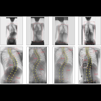
 Results of Cobb angle measurements using the proposed method. The upper row shows the detected region of interest (indicated by a rectangle on the image) and the lower row shows the Cobb angle measurement results. Each column shows examples of (a) standing, (b) supine, (c) bending, and (d) wearing-brace x-ray images. Image courtesy of Scientific Reports through CC BY 4.0.
Three curves were evaluated at the upper, middle, and lower parts of the spine, which they classified as major (largest), minor 1 (next largest), and minor 2 (smallest) curves. Reliability was determined by calculating differences in the Cobb angle measurements by the doctors and the AI using intraclass correlation coefficients (ICCs).
The average Cobb angle difference ranged from approximately from 2.8° to 4.6°, with the largest difference between the doctors' manual average Cobb angle and the AI average Cobb angle being 4.6° at the minor 2 region with patients in a bending position, according to the findings.
Overall, the ICCs recorded for the six doctors and AI were excellent or good, with a value of 0.973 for the major curve in the standing position, the researchers wrote.
"The mean error between AI and doctors was not affected by the angle size, with AI tending to measure 1.7°-2.2° smaller than that measured by the doctors," the authors wrote.
Ultimately, since AIS progresses gradually with growth, it is important not to miss the curve in the early stages of the disease, the researchers noted. Therefore, the ability to detect even small angles may be useful in screening for AIS, they wrote.
Moreover, the total computation time for the AI is a few seconds per case and this could save time and reduce the burden on physicians in medical settings where a huge number of scoliosis measurements are required, according to the researchers.
"The proposed method showed excellent reliability, indicating that it is a promising automated method for measuring [Cobb angle] in patients with AIS," the group concluded.
The full study can be found here.


![A normal mammogram confirmed by three-year radiologic follow-up illustrates reader-marked regions of interest (ROIs) during (A) unaided (round 1) and (B) artificial intelligence (AI)–assisted (round 2) reading. Each colored dot represents an ROI for recall by a human reader. Readers could mark more than one ROI per case, represented by multiple dots of the same color. During AI-assisted reading, the AI system displayed three visible prompts: two with suspicion of malignancy scores of 35% (left mediolateral oblique [L MLO] and craniocaudal [L CC]) and one with a suspicion of malignancy score of 10% (right craniocaudal [R CC]), shown as polygonal overlays. Without AI, six of 10 readers (60%) marked a false-positive ROI. With AI assistance, this fell to two of 10 (20%). R MLO = right mediolateral oblique.](https://img.auntminnie.com/mindful/smg/workspaces/default/uploads/2026/07/2026-07-14-radiology-mammogram-ai-auto-bias.H0bYO8QlWs.jpg?auto=format%2Ccompress&dpr=2&fit=crop&h=167&q=70&w=250)


