Ultrasound with a shear-wave elastography (SWE) technique was comparable to MRI for preoperative evaluation of rotator cuff tears, according to a Thursday presentation at the RSNA 2020 virtual meeting. Elastography measurements showed moderate correlation with MR metrics in the 60-patient study.
Assessing muscle quality is critical for planning surgery to repair the supraspinatus muscle in patients with a torn rotator cuff. While MRI has been long used to evaluate rotator cuff muscles, SWE has emerged as a promising new metric that can be performed at the point of care in order to measure muscle elasticity.
"SWE may be useful to predict tendon repairability by evaluating muscle quality," said presenter Dr. Eun Kyung Khil from the radiology department at Hallym University Dongtan Sacred Heart Hospital in Hwaseong, South Korea.
The prospective study compared ultrasound SWE measurements and conventional MRI metrics for predicting whether surgery to repair the supraspinatus muscle would be successful. It included 60 patients with supraspinatus tears who underwent both preoperative MRI and ultrasound scans between May 2019 and August 2020.
One radiologist with five years of musculoskeletal imaging experience performed the ultrasound scans, which the researchers used to calculate the mean elasticity score, median elasticity score, and elasticity ratio.
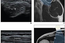
Elasticity was calculated using a longitudinal ultrasound scan of three regions of interest of the supraspinatus muscle, and the scan was repeated three times in order to have nine total region-of-interest measurements. Meanwhile, the elasticity ratio was calculated by dividing the mean elasticity of the supraspinatus muscle by mean elasticity of the trapezius muscle.
In addition, two radiologists read MRI images, which were acquired with a 3-tesla system. The researchers used the following three standard tools to measure muscle evaluation:
- Goutallier grade system to account for fat-to-muscle ratio
- Occupation ratio of the area of supraspinatus muscle to the supraspinatus fossa
- Warner's muscle atrophy grade
The authors compared the ultrasound and MRI measurements for patients whose surgery was successful, defined as a complete or near-complete repair of the rotator cuff, to patients with an incomplete rotator cuff repair.
| MRI and SWE measurements in patients who underwent supraspinatus repair surgery | ||||
| Complete repair | Incomplete repair | p-value | ||
| MRI | Goutallier grade | 1.8 | 3.78 | < 0.001 |
| Occupation ratio | 59.88 | 31.56 | < 0.001 | |
| Muscle atrophy grade | 0.39 | 2.33 | < 0.001 | |
| SWE | Emean, kPa | 31.25 | 43.84 | < 0.001 |
| Emedian, kPa | 29.9 | 43.54 | < 0.001 | |
| Eratio (SST/Tra) | 1.84 | 3.68 | < 0.001 | |
Patients with a successful rotator cuff repair surgery had significantly higher scores on both ultrasound and MRI. Both the mean and median elastography measurements were at least 10 kPa higher in the incomplete repair group.
In addition, Khil said the sensitivity and specificity of SWE were high when using a mean elasticity cutoff value of 35.06 kPa and an elasticity ratio cutoff value of 2.61.
Furthermore, the three elastography measurements on ultrasound showed decent correlation with the MRI metrics. The correlation was particularly strong for the elasticity ratio, which had a coefficient agreement of 0.57 with Goutallier grade and 0.66 with muscle atrophy grade on MRI.
"The correlation coefficient was over 0.4, showing a significant moderate correlation, especially in elasticity ratio," Khil said.
The findings were limited by a small number of participants, especially those in the failed repair group. However, it still demonstrated that SWE looks promising for preoperative evaluation of rotator cuff tears.
"In the preoperative evaluation of [supraspinatus] muscle quality using SWE, especially elasticity ratio, showed moderate correlation with existing MR measurements," Khil said.



![Examples of ultrasound findings and techniques. (A) Images in a 39-year-old male patient with a mass in the left thigh. The mass is heterogeneous on the B-mode US image (compared with the patient in D) and showed increased microvascularity (superb microvascular imaging [SMI]) and shear-wave elastography (SWE) values. Undifferentiated pleomorphic sarcoma was diagnosed at biopsy (with pleomorphic rhabdomyosarcoma in surgical specimen). (B) Images in an 18-year-old male patient with a mass in the left leg. The mass is hypoechoic on the B-mode image, with no other findings suggestive of malignancy. The lesion is in contact with the cortex of the tibia, which is slightly irregular. CT revealed a doubtful anteromedial tibial erosion. The microvascular study demonstrated high vascularization, suggestive of malignancy. Periosteal Ewing sarcoma was diagnosed with both histologic and immunohistochemical confirmation. (C) Images in a 69-year-old female patient with a lump growing on the outside of the left leg. Multiple SWE examinations were performed (please note the high values obtained in the measurements, whereas the color map highlights the stiffness relative to adjacent tissues). SMI showed areas of increased vascularization to target for sampling. Undifferentiated spindle cell sarcoma was diagnosed at biopsy, with residual leiomyosarcoma in the surgical specimen after neoadjuvant therapy. (D) Images in a 56-year-old female patient with a mass in the right thigh. The mass is heterogeneous at both B-mode ultrasound (similar to patient A) and MRI (coronal T2-weighted spectral attenuated inversion recovery [SPAIR]; T1-weighted pre-contrast and postcontrast imaging), which even shows uptake after the administration of paramagnetic contrast material, which is traditionally suggestive of malignancy. Low values at SMI and elastography are suggestive of benignity. Spindle cell lipoma was diagnosed at biopsy, with atypical spindle cell lipomatous tumor in the surgical specimen.](https://img.auntminnie.com/mindful/smg/workspaces/default/uploads/2026/08/images-radiol250278fig2.APCFLSvX6p.jpg?auto=format%2Ccompress&fit=crop&h=100&q=70&w=100)


![Examples of ultrasound findings and techniques. (A) Images in a 39-year-old male patient with a mass in the left thigh. The mass is heterogeneous on the B-mode US image (compared with the patient in D) and showed increased microvascularity (superb microvascular imaging [SMI]) and shear-wave elastography (SWE) values. Undifferentiated pleomorphic sarcoma was diagnosed at biopsy (with pleomorphic rhabdomyosarcoma in surgical specimen). (B) Images in an 18-year-old male patient with a mass in the left leg. The mass is hypoechoic on the B-mode image, with no other findings suggestive of malignancy. The lesion is in contact with the cortex of the tibia, which is slightly irregular. CT revealed a doubtful anteromedial tibial erosion. The microvascular study demonstrated high vascularization, suggestive of malignancy. Periosteal Ewing sarcoma was diagnosed with both histologic and immunohistochemical confirmation. (C) Images in a 69-year-old female patient with a lump growing on the outside of the left leg. Multiple SWE examinations were performed (please note the high values obtained in the measurements, whereas the color map highlights the stiffness relative to adjacent tissues). SMI showed areas of increased vascularization to target for sampling. Undifferentiated spindle cell sarcoma was diagnosed at biopsy, with residual leiomyosarcoma in the surgical specimen after neoadjuvant therapy. (D) Images in a 56-year-old female patient with a mass in the right thigh. The mass is heterogeneous at both B-mode ultrasound (similar to patient A) and MRI (coronal T2-weighted spectral attenuated inversion recovery [SPAIR]; T1-weighted pre-contrast and postcontrast imaging), which even shows uptake after the administration of paramagnetic contrast material, which is traditionally suggestive of malignancy. Low values at SMI and elastography are suggestive of benignity. Spindle cell lipoma was diagnosed at biopsy, with atypical spindle cell lipomatous tumor in the surgical specimen.](https://img.auntminnie.com/mindful/smg/workspaces/default/uploads/2026/08/images-radiol250278fig2.APCFLSvX6p.jpg?auto=format%2Ccompress&fit=crop&h=112&q=70&w=112)