Handheld ultrasound scanners are invaluable tools for performing imaging in patients with COVID-19, according to a Wednesday presentation at the RSNA 2020 virtual meeting.
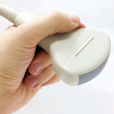
Presenter Zhili Xu acknowledged that CT is the optimal imaging modality for patients with COVID-19, but she hoped radiologists would also consider adding ultrasound to their pandemic imaging arsenal. Notably, handheld ultrasound machines are easy to clean, transport, and disinfect -- crucial benefits during an infectious disease pandemic.
"Ultrasound machines have become more and more portable," said Xu from Guangdong Second Provincial General Hospital in China. "Especially wireless handheld ultrasound is very suitable for COVID-19-infected patients in isolation wards."
COVID-19 was first documented in Wuhan, China, in late 2019. Since then, the World Health Organization declared the disease a pandemic, with more than 37 million cumulative cases and 1 million total deaths.
To help address a surge of patients in Wuhan, the Chinese government built temporary mobile cabin hospitals and a tent hospital. Xu and her colleagues used wireless ultrasound machines to scan patients at one of the temporary mobile cabin locations.
Xu and her team have primarily used wireless handheld devices to look for abdominal disease in COVID-19 patients. They used ultrasound to diagnose patients with numerous conditions, including cholecystolithiasis, renal calculus with hydronephrosis, and gouty arthritis.
Although it wasn't their main focus, Xu and colleagues also used wireless ultrasound machines to diagnose and monitor pneumonia in two patients, adding to the list of numerous researchers who have used ultrasound for pulmonary imaging during the pandemic.
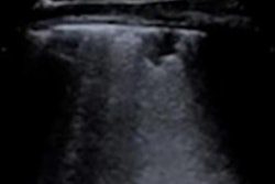
For lung imaging, the team utilized the Bedside Lung Ultrasound in Emergency (BLUE) protocol, which uses a physician's hands to guide probe placement. Like other teams, they looked for hallmark signs of COVID-19 on ultrasound, including B-lines and lung consolidation.
"Three B-lines or more between two ribs demonstrate interstitial syndrome, which is a common thing in COVID-19 patients," Xu said.
While patients become infected with COVID-19 through respiratory or aerosol transmission, Xu also said it would be a mistake to ignore the risk of contact transmission. This is where portable ultrasound machines really shine.
"For the advantages of convenience, security, price, and real-time imaging, ultrasound was widely used in [our] clinic," she said.
Xu recommended taking a three-step infection control approach for preparing ultrasound machines for COVID-19 isolation wards:
- Wipe the screen and probe with an antifog cloth.
- Spread couplant gel on the probe.
- Wrap both the screen and probe in two layers of protective film.
Once patients have been scanned, carefully remove the protective film, then spray the probe and device with 75% ethyl alcohol, she advised.
Despite its many advantages during an infectious disease pandemic, ultrasound is not perfect. The modality cannot compete with CT for sensitive lung imaging, and it suffers from poor spatial resolution -- an area Xu said needs more technical improvements.
However, even with its drawbacks, ultrasound has an unrivaled ability to safely, conveniently, and cheaply scan patients with COVID-19, according to Xu.
"Wireless handheld ultrasound contributes to quick diagnosis of complications and reduced contact transmission of COVID-19, which will increase the cure rate of COVID-19 infection and delay the progress of this epidemic," she concluded.

![Examples of ultrasound findings and techniques. (A) Images in a 39-year-old male patient with a mass in the left thigh. The mass is heterogeneous on the B-mode US image (compared with the patient in D) and showed increased microvascularity (superb microvascular imaging [SMI]) and shear-wave elastography (SWE) values. Undifferentiated pleomorphic sarcoma was diagnosed at biopsy (with pleomorphic rhabdomyosarcoma in surgical specimen). (B) Images in an 18-year-old male patient with a mass in the left leg. The mass is hypoechoic on the B-mode image, with no other findings suggestive of malignancy. The lesion is in contact with the cortex of the tibia, which is slightly irregular. CT revealed a doubtful anteromedial tibial erosion. The microvascular study demonstrated high vascularization, suggestive of malignancy. Periosteal Ewing sarcoma was diagnosed with both histologic and immunohistochemical confirmation. (C) Images in a 69-year-old female patient with a lump growing on the outside of the left leg. Multiple SWE examinations were performed (please note the high values obtained in the measurements, whereas the color map highlights the stiffness relative to adjacent tissues). SMI showed areas of increased vascularization to target for sampling. Undifferentiated spindle cell sarcoma was diagnosed at biopsy, with residual leiomyosarcoma in the surgical specimen after neoadjuvant therapy. (D) Images in a 56-year-old female patient with a mass in the right thigh. The mass is heterogeneous at both B-mode ultrasound (similar to patient A) and MRI (coronal T2-weighted spectral attenuated inversion recovery [SPAIR]; T1-weighted pre-contrast and postcontrast imaging), which even shows uptake after the administration of paramagnetic contrast material, which is traditionally suggestive of malignancy. Low values at SMI and elastography are suggestive of benignity. Spindle cell lipoma was diagnosed at biopsy, with atypical spindle cell lipomatous tumor in the surgical specimen.](https://img.auntminnie.com/mindful/smg/workspaces/default/uploads/2026/08/images-radiol250278fig2.APCFLSvX6p.jpg?auto=format%2Ccompress&fit=crop&h=100&q=70&w=100)


![Examples of ultrasound findings and techniques. (A) Images in a 39-year-old male patient with a mass in the left thigh. The mass is heterogeneous on the B-mode US image (compared with the patient in D) and showed increased microvascularity (superb microvascular imaging [SMI]) and shear-wave elastography (SWE) values. Undifferentiated pleomorphic sarcoma was diagnosed at biopsy (with pleomorphic rhabdomyosarcoma in surgical specimen). (B) Images in an 18-year-old male patient with a mass in the left leg. The mass is hypoechoic on the B-mode image, with no other findings suggestive of malignancy. The lesion is in contact with the cortex of the tibia, which is slightly irregular. CT revealed a doubtful anteromedial tibial erosion. The microvascular study demonstrated high vascularization, suggestive of malignancy. Periosteal Ewing sarcoma was diagnosed with both histologic and immunohistochemical confirmation. (C) Images in a 69-year-old female patient with a lump growing on the outside of the left leg. Multiple SWE examinations were performed (please note the high values obtained in the measurements, whereas the color map highlights the stiffness relative to adjacent tissues). SMI showed areas of increased vascularization to target for sampling. Undifferentiated spindle cell sarcoma was diagnosed at biopsy, with residual leiomyosarcoma in the surgical specimen after neoadjuvant therapy. (D) Images in a 56-year-old female patient with a mass in the right thigh. The mass is heterogeneous at both B-mode ultrasound (similar to patient A) and MRI (coronal T2-weighted spectral attenuated inversion recovery [SPAIR]; T1-weighted pre-contrast and postcontrast imaging), which even shows uptake after the administration of paramagnetic contrast material, which is traditionally suggestive of malignancy. Low values at SMI and elastography are suggestive of benignity. Spindle cell lipoma was diagnosed at biopsy, with atypical spindle cell lipomatous tumor in the surgical specimen.](https://img.auntminnie.com/mindful/smg/workspaces/default/uploads/2026/08/images-radiol250278fig2.APCFLSvX6p.jpg?auto=format%2Ccompress&fit=crop&h=112&q=70&w=112)