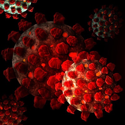
The American Institute of Ultrasound in Medicine (AIUM) has changed its ultrasound cleaning and safe handling guidelines amid the ongoing novel coronavirus outbreak.
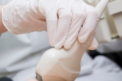
The updated guidelines specify that healthcare professionals can disinfect external and interventional transducers with soap and water if low-level disinfection materials are not available. In addition, medical gloves, medical dressings, and other physical barriers should be used if there are no transducer covers.
The AIUM also noted that low-level disinfection is effective for external and interventional procedures, according to the U.S. Centers for Disease Control and Prevention. The guidelines include a link to disinfectants approved by the U.S. Environmental Protection Agency for use against the novel coronavirus.
Other changes to the guidelines for the COVID-19 pandemic include the following:
- Equipment requires full low-level disinfection if used for patients with COVID-19 and other respiratory infections that require mechanical ventilation, aerosolization application, and other aerosolization procedures.
- If possible, use a single, dedicated scanner and transducer system for patients with suspected or confirmed cases of COVID-19, as the novel coronavirus can survive on plastic for up to 72 hours.
- Clean all ancillary equipment for a procedure when cleaning an ultrasound system.
- Follow manufacturer and institutional guidelines.







![Examples of ultrasound findings and techniques. (A) Images in a 39-year-old male patient with a mass in the left thigh. The mass is heterogeneous on the B-mode US image (compared with the patient in D) and showed increased microvascularity (superb microvascular imaging [SMI]) and shear-wave elastography (SWE) values. Undifferentiated pleomorphic sarcoma was diagnosed at biopsy (with pleomorphic rhabdomyosarcoma in surgical specimen). (B) Images in an 18-year-old male patient with a mass in the left leg. The mass is hypoechoic on the B-mode image, with no other findings suggestive of malignancy. The lesion is in contact with the cortex of the tibia, which is slightly irregular. CT revealed a doubtful anteromedial tibial erosion. The microvascular study demonstrated high vascularization, suggestive of malignancy. Periosteal Ewing sarcoma was diagnosed with both histologic and immunohistochemical confirmation. (C) Images in a 69-year-old female patient with a lump growing on the outside of the left leg. Multiple SWE examinations were performed (please note the high values obtained in the measurements, whereas the color map highlights the stiffness relative to adjacent tissues). SMI showed areas of increased vascularization to target for sampling. Undifferentiated spindle cell sarcoma was diagnosed at biopsy, with residual leiomyosarcoma in the surgical specimen after neoadjuvant therapy. (D) Images in a 56-year-old female patient with a mass in the right thigh. The mass is heterogeneous at both B-mode ultrasound (similar to patient A) and MRI (coronal T2-weighted spectral attenuated inversion recovery [SPAIR]; T1-weighted pre-contrast and postcontrast imaging), which even shows uptake after the administration of paramagnetic contrast material, which is traditionally suggestive of malignancy. Low values at SMI and elastography are suggestive of benignity. Spindle cell lipoma was diagnosed at biopsy, with atypical spindle cell lipomatous tumor in the surgical specimen.](https://img.auntminnie.com/mindful/smg/workspaces/default/uploads/2026/08/images-radiol250278fig2.APCFLSvX6p.jpg?auto=format%2Ccompress&fit=crop&h=112&q=70&w=112)


