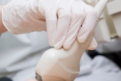
Ultrasound has played a key role in the intensive care unit (ICU) during the COVID-19 pandemic, according to a panel held April 13 at the virtual 2021 American Institute of Ultrasound in Medicine (AIUM) annual meeting.
That being said, going forward there's still a need to establish clear protocols and training for the use of the modality in a health crisis, especially since operator skill can vary, panelist Dr. David Schrift of Prisma Health Midlands/University of South Carolina School of Medicine Columbia told session attendees.
Schrift was joined on the panel by Dr. Michael Lanspa of the University of Utah in Salt Lake City. The talk was moderated by Dr. Rachel Liu of Yale University School of Medicine in New Haven, CT, and Dr. Keith Barron, also of Prisma Health Midlands.
In addition to addressing ultrasound protocols, Schrift and Lanspa discussed the most useful ultrasound applications during the peak of the COVID-19 pandemic.
Ultrasound has proven to be a useful modality during the pandemic in helping clinicians determine factors that contribute to respiratory failure in patients, with the most common exams being cardiac and lung, Schrift said.
"[The patients we were evaluating were already diagnosed with] COVID-19, so the question is, what are the factors contributing to respiratory failure?" he said.
By the time patients were admitted to the ICU, imaging was less about diagnosis and more about deciding on and managing treatment, Lanspa said.
"[We found ultrasound in the ICU to be] most useful in managing patients who had reached the limits of conventional therapy," he said.
However, both Schrift and Lanspa agreed that at times ultrasound was not the ideal imaging modality for COVID-19 patients -- in part because its use could expose more healthcare staff to infection.
"There are a lot of things you can do with ultrasound, but specifically for COVID-19 there are times it's not helpful, particularly the peri-intubation time point," Schrift said. "The reality is that a single x-ray in those instances is very high yield: It takes very little time, there's only one person in the room, you don't need the second person you tend to need to confirm the tube with ultrasound."
In response to a question from the AIUM moderators about how ultrasound can be used effectively in other health crises in the future, both Schrift and Lanspa agreed that establishing clear protocols for ultrasound in a COVID-19 era is important. And an effective ultrasound protocol for a disease like COVID-19 should take into consideration not only presentation and progression of patients but also what kinds of care teams are necessary, Lanspa noted.
"[Back in March and April,] I was involved in a lot of discussion [about what we were doing to cope with COVID-19]," he said. "People pitched protocols, and all were very different ... designed for a certain type of practitioner, a certain level of knowledge, a certain environment ... What we need to tackle is [establishing protocols that provide a] level of training we expect everyone to have."
In the end, COVID-19 often manifests as acute respiratory distress syndrome (ARDS) and can be handled accordingly, Lanspa said.
"We watched an entire year of people rediscovering how to treat ARDS as if it was a brand-new thing," he said. "What we need is a robust protocol designed for severe ARDS patient ... Who knows how long COVID will last? But ARDS will persist."