Ultrasound experts released guidelines for performing safe scans during the ongoing COVID-19 crisis. The guidelines, which were published on April 4 in Ultrasound in Medicine and Biology, reinforce standard cleaning and disinfection protocols, as well as introduce new measures in light of the pandemic.
The advice comes from an infectious disease expert and ob/gyn doctor on behalf of the World Federation for Ultrasound in Medicine and Biology Safety Committee. While the authors know that not every guideline will apply to all clinicians, they hope the guidance contributes to the safety practitioners and patients.
Some doctors have been using ultrasound systems to perform lung sonography on patients with COVID-19. While ultrasound equipment has multiple benefits, including use at the bedsides of patients, precautions still must be taken to ensure the safety of clinicians, staff, and patients.
The guidelines detail five domains of recommendations for ultrasound safety during the ongoing pandemic.
1. Socially distance patients
Healthcare settings performing ultrasound scans should be mindful of social distancing for patient safety. This includes scheduling appointments at infrequent intervals, spacing seats in waiting rooms at least six feet apart, and being mindful of the time during scheduled visits.
Practitioners also should ideally limit visitors to a maximum of one person, the guidelines noted. However, video visits or phone calls could be a way to virtually bring additional visitors to appointments.
2. Use PPE
Personal protective equipment (PPE) is vital to protect clinicians and staff during the ongoing pandemic.
Clinicians and staff should use a surgical face mask before entering a patient room or care area, the authors noted. In addition, respirator masks, such as N95 masks, should be used for procedures that generate aerosol.
Gloves and associated hand hygiene are also essential for professionals who use ultrasound equipment. Ultrasound staff should perform adequate hand hygiene before and after patient visits, as well as after touching potentially infectious surfaces or removing PPE.
Clinicians who are at increased health risk due to the pandemic should not perform ultrasound procedures, the authors noted.
3. Prepare the room
At the beginning of the day, the ultrasound room should be cleaned by wiping all surfaces and equipment with a low-level disinfectant. The authors also advised removing unnecessary accessories and replacing fabric chairs with ones that can be easily wiped down with disinfectant.
Between patient visits, staff should wipe down the bed or couch. They should also use gloves to fold and dispose of paper covers.
If possible, the authors recommended scanning patients with suspected or confirmed cases of COVID-19 at the end of the day. This allows extra time for vigorous cleaning and disinfection, they noted.
4. Disinfect and clean the equipment
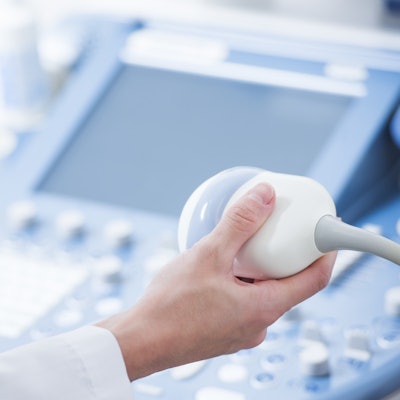
Ultrasound transducers and cables should be cleaned and disinfected between patients.
Transducers used for lung ultrasound scans can be cleaned with a low- to intermediate-level disinfectant, the authors noted. Staff should also wear gloves and perform hand hygiene when cleaning and disinfecting equipment.
The authors recommended dedicating at least one ultrasound machine specifically for patients with suspected or confirmed cases of COVID-19. Further, clinicians should keep the minimum number of transducers needed out and available, while storing the rest behind cabinet doors
5. Use single-use gel packets
Finally, in light of the pandemic, the authors recommended single-use, nonsterile gel packets for external ultrasound examinations on patients with confirmed or suspected cases of COVID-19.
When gel packets are not available, the authors advised clinicians to use gel bottles for external exams only. However, these bottles should not be refilled or heated, they noted.
The authors further recommended that clinicians keep one hand clean for patient interactions while using their other "semi-clean" hand to dispense gel and perform other nonpatient tasks, such as typing.