Using artificial intelligence (AI) with thyroid ultrasound offers a quick and noninvasive approach to thyroid cancer screening, according to a study published online on October 24 in JAMA Otolaryngology -- Head & Neck Surgery.
The findings suggest that clinicians may have a better way for distinguishing between low- and high-risk thyroid nodules, since methods such as biopsy can be inconclusive, said study co-author Dr. Elizabeth Cottrill from Thomas Jefferson University in Philadelphia, in a statement.
"Currently, ultrasounds can tell us if a nodule looks suspicious, and then the decision is made whether to do a needle biopsy or not," Cottrill said. "But fine-needle biopsies only act as a peephole -- they don't tell us the whole picture. As a result, some biopsies return inconclusive results for whether or not the nodule may be malignant."
The prevalence of thyroid nodules in the general population is as high as 67%, the authors noted. Most nodules are benign, but once one is detected, it must be evaluated via ultrasound. Nodules larger than 1 cm are often recommended for fine-needle biopsy; some are evaluated with molecular genetic testing, which, although less invasive, is expensive.
"Machine learning is a low-cost and efficient tool that could help physicians arrive at a quicker decision as to how to approach an indeterminate nodule," said senior author John Eisenbrey, PhD, also of Thomas Jefferson University, in the statement.
The researchers investigated whether a machine-learning algorithm developed on a Google platform could be used with thyroid ultrasound to serve as a quick and inexpensive first screen for thyroid cancer. They trained the algorithm on 556 thyroid nodule images from 121 patients who underwent ultrasound-guided fine-needle biopsy and, subsequently, molecular testing.
Based on a particular gene panel, the molecular testing categorized 43 of the nodules as high risk and 91 as low risk. The group then tested the algorithm on a different set of 53 images to determine how well it could classify thyroid nodules compared with molecular test results.
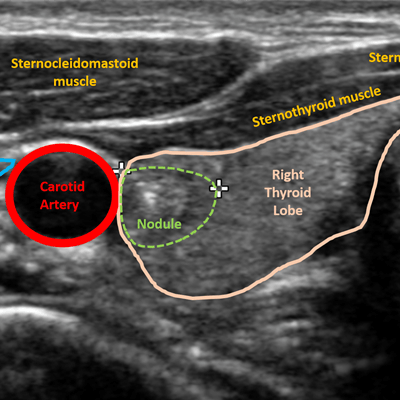
 Ultrasound image of thyroid nodule. Image courtesy of Dr. Elizabeth Cottrill, Thomas Jefferson University.
The team found that the algorithm's specificity was high -- an indicator of a low rate of false positives -- as was its positive predictive value (PPV). Its overall accuracy was 77.4%.
| AI algorithm's performance classifying thyroid nodules | |
| Performance measure | Performance |
| Specificity | 97% |
| Positive predictive value | 90% |
| Accuracy | 77.4% |
| Negative predictive value | 74.4% |
| Sensitivity | 45% |
"The model we built performed with a high specificity (97%) and PPV (90%) for ruling in the presence of a high-risk mutation on the basis of sonography alone," the group noted.
Although the study results are preliminary, they imply that AI could be a promising tool for improving the diagnosis of thyroid cancer.
"There are so many potential applications of machine learning," Eisenbrey said. "In the future, we'd like to make use of feature extraction, which will help us identify anatomically relevant features of high-risk nodules."