Thyroid cancer diagnoses are up to three times more common in 9/11 first responders than the general population. However, the increased cancer rate may be due to overscreening, according to an article published on April 20 in JAMA Internal Medicine.
Firefighters and emergency medical personnel who responded to the September 11 attacks at the World Trade Center (WTC) were likely exposed to a huge amount of various toxins. Since the attack, these first responders have received thorough annual medical monitoring that includes CT, MRI, and PET imaging.
But the authors believe these regular exams account for the higher rate of thyroid cancer among first responders, rather than exposure to toxins.
"The elevated thyroid cancer rate may ... be an artifact of such frequent and detailed medical surveillance," wrote the authors, led by Hilary Colbeth from the Fire Department of the City of New York and Montefiore Medical Center.
The study included data from 14,987 male firefighters and emergency medical personnel on the scene during the 9/11 World Trade Center attack. These first responders participated in the WTC Health Program, in which they were eligible to receive detailed medical screenings every 12-18 months that included chest CT scans and head and neck MRI.
Between 2001 and 2019, 72 first responders in the screening program developed thyroid cancer. One person was diagnosed with follicular carcinoma, while the remaining 98% of cases were papillary thyroid cancer. None of the first responders died as a result of their cancer, and none developed metastatic tumors.
The authors then compared the thyroid cancer rates of the first responders with a similar cohort of men from Olmsted County, Minnesota.
At first, the researchers found no difference in thyroid cancer incidence between the two groups. But thyroid cancer diagnoses for first responders increased after Congress passed the James L. Zadroga 9/11 Health and Compensation Act, which improved access to care for first responders, the authors noted.
The first responders had twice the thyroid cancer incidence rate as the men in Olmsted County. They were also more likely than their Minnesota peers to be asymptomatic (81.5% versus 54.5%).
"Given that detection of subclinical pools of thyroid cancer can be directly related to healthcare access, we believe the totality of our results suggests that incidental detection has played a role in the elevated incidence of WTC-exposed thyroid cancers reported by previous studies," the authors wrote.
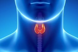
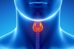
Furthermore, the authors believed it was likely many first responders with thyroid cancer received a lobectomy or total thyroidectomy, a costly treatment that may not be needed for all patients. Instead, it may be better for clinicians to take a wait-and-see approach for 9/11 first responders.
"One strategy to attenuate a high thyroid cancer diagnosis rate was enacted by South Korea, which began to conduct fewer screenings in March 2014 and since then has experienced fewer diagnosis and surgical operations," the authors wrote. "Alternatively, the American Thyroid Association has recommended adopting an active surveillance program for low-risk papillary microcarcinomas, which could be a treatment option for qualifying WTC-exposed patients."

![Examples of ultrasound findings and techniques. (A) Images in a 39-year-old male patient with a mass in the left thigh. The mass is heterogeneous on the B-mode US image (compared with the patient in D) and showed increased microvascularity (superb microvascular imaging [SMI]) and shear-wave elastography (SWE) values. Undifferentiated pleomorphic sarcoma was diagnosed at biopsy (with pleomorphic rhabdomyosarcoma in surgical specimen). (B) Images in an 18-year-old male patient with a mass in the left leg. The mass is hypoechoic on the B-mode image, with no other findings suggestive of malignancy. The lesion is in contact with the cortex of the tibia, which is slightly irregular. CT revealed a doubtful anteromedial tibial erosion. The microvascular study demonstrated high vascularization, suggestive of malignancy. Periosteal Ewing sarcoma was diagnosed with both histologic and immunohistochemical confirmation. (C) Images in a 69-year-old female patient with a lump growing on the outside of the left leg. Multiple SWE examinations were performed (please note the high values obtained in the measurements, whereas the color map highlights the stiffness relative to adjacent tissues). SMI showed areas of increased vascularization to target for sampling. Undifferentiated spindle cell sarcoma was diagnosed at biopsy, with residual leiomyosarcoma in the surgical specimen after neoadjuvant therapy. (D) Images in a 56-year-old female patient with a mass in the right thigh. The mass is heterogeneous at both B-mode ultrasound (similar to patient A) and MRI (coronal T2-weighted spectral attenuated inversion recovery [SPAIR]; T1-weighted pre-contrast and postcontrast imaging), which even shows uptake after the administration of paramagnetic contrast material, which is traditionally suggestive of malignancy. Low values at SMI and elastography are suggestive of benignity. Spindle cell lipoma was diagnosed at biopsy, with atypical spindle cell lipomatous tumor in the surgical specimen.](https://img.auntminnie.com/mindful/smg/workspaces/default/uploads/2026/08/images-radiol250278fig2.APCFLSvX6p.jpg?auto=format%2Ccompress&fit=crop&h=100&q=70&w=100)


![Examples of ultrasound findings and techniques. (A) Images in a 39-year-old male patient with a mass in the left thigh. The mass is heterogeneous on the B-mode US image (compared with the patient in D) and showed increased microvascularity (superb microvascular imaging [SMI]) and shear-wave elastography (SWE) values. Undifferentiated pleomorphic sarcoma was diagnosed at biopsy (with pleomorphic rhabdomyosarcoma in surgical specimen). (B) Images in an 18-year-old male patient with a mass in the left leg. The mass is hypoechoic on the B-mode image, with no other findings suggestive of malignancy. The lesion is in contact with the cortex of the tibia, which is slightly irregular. CT revealed a doubtful anteromedial tibial erosion. The microvascular study demonstrated high vascularization, suggestive of malignancy. Periosteal Ewing sarcoma was diagnosed with both histologic and immunohistochemical confirmation. (C) Images in a 69-year-old female patient with a lump growing on the outside of the left leg. Multiple SWE examinations were performed (please note the high values obtained in the measurements, whereas the color map highlights the stiffness relative to adjacent tissues). SMI showed areas of increased vascularization to target for sampling. Undifferentiated spindle cell sarcoma was diagnosed at biopsy, with residual leiomyosarcoma in the surgical specimen after neoadjuvant therapy. (D) Images in a 56-year-old female patient with a mass in the right thigh. The mass is heterogeneous at both B-mode ultrasound (similar to patient A) and MRI (coronal T2-weighted spectral attenuated inversion recovery [SPAIR]; T1-weighted pre-contrast and postcontrast imaging), which even shows uptake after the administration of paramagnetic contrast material, which is traditionally suggestive of malignancy. Low values at SMI and elastography are suggestive of benignity. Spindle cell lipoma was diagnosed at biopsy, with atypical spindle cell lipomatous tumor in the surgical specimen.](https://img.auntminnie.com/mindful/smg/workspaces/default/uploads/2026/08/images-radiol250278fig2.APCFLSvX6p.jpg?auto=format%2Ccompress&fit=crop&h=112&q=70&w=112)