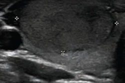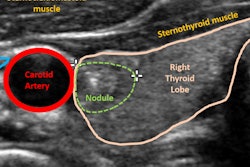
Radiofrequency ultrasound and an artificial intelligence (AI) model can be used to effectively predict the malignancy of thyroid nodules, as well as stratify their risk, according to a study set for publication in the November issue of Ultrasonics.
The combination of radiofrequency ultrasound with an artificial neural network (ANN) could also avoid operator dependency issues and help prevent unnecessary thyroid biopsies, according to a group led by Dr. Chunrui Liu of the Affiliated Hospital of Nanjing University Medical School in China.
"The proposed method has no operator dependency; all of the analyses are performed by computer," the team noted (Ultrasonics, November 2019, Vol. 99, pp. 1-9). "Preliminary results indicated that the performance of ANN combined with radiofrequency ultrasound signals is better than that combined with conventional ultrasound images."
Common but not often malignant
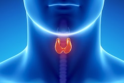
Thyroid nodules are common, but only 8% to 16% are actually malignant, according to the researchers. Many ultrasound techniques are used to evaluate nodule malignancy, including strain elastography, acoustic radiation force impulse imaging, and contrast-enhanced ultrasound, but these methods' efficacy remains unclear, the group wrote.
That's where radiofrequency ultrasound comes in. The technique elicits more clinical information than conventional ultrasound by extracting radiofrequency signals from tissues. But how it performs with thyroid nodules has not been studied, Liu and colleagues noted.
"Preliminary studies of radiofrequency ultrasound have been promising, and the method has been shown to have broader prospective applications in identifying prostate and breast cancers and grading fatty liver," they wrote. "To date, few studies on radiofrequency ultrasound's thyroid cancer detection performance have been reported."
The researchers developed their method to predict suspicious thyroid nodules by first gathering radiofrequency data and then creating radiofrequency ultrasound images using Matlab software (MathWorks). After a radiologist outlined regions of interest on the images, textural features were then analyzed using the gray-level co-occurrence matrix (GLCM) algorithm and principal component analysis. The resulting characteristic values from the textural analysis were subsequently used to train the ANN.
The study included 131 pathologically proven thyroid nodules, of which 59 were benign and 72 were malignant. The nodules were randomly divided into training, validation, and testing cohorts. To test their hypothesis that radiofrequency ultrasound could provide more tissue characteristic information than conventional ultrasound, the researchers also performed the same texture and ANN analyses on the B-mode ultrasound images.
The ANN algorithm performed better with radiofrequency ultrasound than it did on conventional ultrasound in all categories except specificity, the group found.
| ANN performance for predicting thyroid nodule malignancy | ||
| Performance measure | ANN on conventional ultrasound images | ANN on radiofrequency ultrasound images |
| Sensitivity | 94.4% | 100% |
| Specificity | 93.2% | 91.5% |
| Accuracy | 93.9% | 96.2% |
| AUC* | 0.917 | 0.945 |
The group also used the ANN with radiofrequency ultrasound to characterize new malignancy risk groups for categories 3 (probably benign), 4 (suspicious), and 5 (probably malignant) thyroid nodules as established by the American College of Radiology's Thyroid Imaging Reporting and Data System (TI-RADS). The new categories better distinguished malignant nodules compared with TI-RADS.
| Parameter | Category 3 | Category 4 | Category 5 |
| No. of samples | 42 | 16 | 73 |
| No. of malignant samples | 0 | 3 | 69 |
| Risk of malignancy | |||
| ANN plus radiofrequency ultrasound | 0 | 18.8% | 94.5% |
| TI-RADS | 0 | 55.1% | 88.2% |
"The new categories allow for a selection of suspicious nodules to be submitted to fine-needle aspiration, thereby avoiding unnecessary thyroid biopsies," the group wrote.
More research to come
More research needs to be done to establish the benefits of using an ANN and radiofrequency ultrasound, according to Liu and colleagues.
"Of course, although these preliminary results suggested [the use of the ANN and radiofrequency ultrasound] could help sonographers to identify risky thyroid nodules and reduce the number of unnecessary thyroid biopsies, more data will be collected and analyzed in our future study to further confirm the feasibility and accuracy of the proposed method," they concluded.