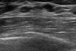
Ultrasound with a shear-wave elastography (SWE) technique offers an effective way to evaluate Achilles tendon health in both athletes and nonathletes, according to a study published online January 14 in Academic Radiology.
Studies have demonstrated that SWE is useful for evaluating tendon stiffness and can aid radiologists in diagnosing tendinopathy better than other ultrasound modes, as diseased or injured tendons are generally softer than healthy ones, wrote a team led by Dr. Timm Dirrichs of RWTH Aachen University Hospital in Germany. But reference data from varying population groups are lacking.
"[Our main] objective ... was to analyze tendon stiffness in semiprofessional long- and middistance running athletes and to compare [their] tendon stiffness to a nonathletic control group," the group wrote. "[The] second objective was to evaluate the specificity with which SWE was able to predict the absence of clinical symptoms."
Tendons are currently imaged primarily with B-mode ultrasound, power Doppler ultrasound, and MRI. But these methods do not always accurately visualize tendinopathy -- or help clinicians track the transition from asymptomatic to symptomatic disease, according to the researchers.
"In up to 59% of asymptomatic patients, especially athletes, abnormal imaging is present in various tendons, while ... B-mode ultrasound and power Doppler ultrasound might remain inconspicuous in patients with clinical relevant tendinopathy," they noted.
That's where SWE comes in, the researchers wrote. It provides quantitative information about tissue stiffness and, therefore, about the mechanical properties of a tendon, which could be used to estimate tendon integrity.
The study included 68 asymptomatic healthy participants: 33 were semiprofessional athletes who completed at least five training sessions of running per week, and 35 were nonathletic individuals. Each participant's Achilles tendons were imaged with B-mode ultrasound, power Doppler ultrasound, and SWE (136 tendons). SWE values were expressed in kilopascals (kPa).
The mean SWE value for the Achilles tendon was 183.8 kPa in athletes and 103.6 kPa in nonathletes, a difference that was statistically significant (p < 0.001).
"We found athletes' Achilles tendons to be 1.8 times as hard as nonathletes' tendons obtained by SWE," the group noted. "This phenomenon may be caused by repeated training effects."
The researchers found that SWE outperformed both B-mode and power Doppler ultrasound in terms of the specificity of classifying an asymptomatic tendon as healthy:
- B-mode ultrasound: 60.6%
- Power Doppler ultrasound: 93.9%
- SWE: 96.3%
SWE is useful for measuring and displaying the effects of training on the Achilles tendons of athletes, and its high specificity could help clinicians better care for patients, the group wrote.
"[SWE] ... has a high specificity to predict absence of clinical symptoms," Dirrichs and colleagues concluded. "This might be of great clinical interest, especially in asymptomatic athletes, where the rate of false-positive findings in conventional B-mode ultrasound and power Doppler ultrasound is high."