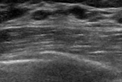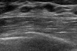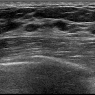
Ultrasound with a shear-wave elastography (SWE) technique can distinguish benign from malignant breast microcalcifications detected on ultrasound, according to a new study published in the August issue of the American Journal of Roentgenology.
The study findings could help physicians better plan a response to microcalcifications, wrote a team led by Dr. Foucauld Chamming's of Institut Bergonié, Bordeaux Cedex, France.
"On SWE, microcalcifications associated with malignant lesions showed significantly higher stiffness values than benign ones," the group wrote. "In the clinical setting, these results are of particular interest for assessment of concordance between radiologic and pathologic findings."
Digital mammography has long been the gold standard for identifying microcalcifications, but its specificity for characterizing them is low -- ranging from 10% to 67% -- leading to a high number of benign biopsies, the authors noted. Suspicious microcalcifications are often worked up with ultrasound to find signs of invasive cancer and any additional lesions, but conventional ultrasound has its limitations, Chamming's and colleagues wrote (AJR, August 2019, Vol. 213:2, pp. W85-W92).
"Although malignant microcalcifications are more likely to be seen on ultrasound than benign ones, B-mode ultrasound does not provide relevant additional information for the characterization of isolated microcalcifications," they wrote. "Shear-wave elastography, which is able to measure tissue stiffness quantitatively, is a relatively recent ultrasound technique and has been shown to improve characterization of breast lesions, especially masses."
Chamming's and colleagues investigated whether SWE could distinguish benign from malignant microcalcifications of the breast by conducting a study that included 74 patients with mammographically detected suspicious microcalcifications who underwent breast ultrasound between February and June 2016. All of the patients also had an SWE exam; those with malignant microcalcifications were biopsied using ultrasound guidance.
The researchers compared qualitative and quantitative elastography results between benign and malignant calcifications, as well as between pure ductal carcinoma in situ (DCIS) and invasive lesions. They used area under the receiver operating characteristic curves (AUC) to evaluate SWE's performance in detecting malignancy and invasive features of breast lesions.
Ultrasound identified 29 groups of microcalcifications in 29 patients. Pathology results showed 16 benign and 13 malignant groups of microcalcifications.
The authors found that the stiffness of malignant calcifications was higher than benign ones (p = 0.0004). SWE had 100% specificity and positive predictive value for both detecting malignant calcifications and identifying invasive features of breast lesions.
| SWE's performance for breast lesions | ||
| Performance measure | Diagnosis of malignancy | Detection of invasive features |
| AUC | 0.89 | 0.93 |
| Sensitivity | 69% | 75% |
| Specificity | 100% | 100% |
| Negative predictive value | 80% | 75% |
| Positive predictive value | 100% | 100% |
| Accuracy | 86% | 85% |
The study results could help physicians better manage microcalcifications, according to the authors.
"When faced with stiff calcifications, which have a high probability of being malignant, a biopsy yielding benign results should be questioned, and a second biopsy or surgical excision should be considered," the group concluded.


![Examples of ultrasound findings and techniques. (A) Images in a 39-year-old male patient with a mass in the left thigh. The mass is heterogeneous on the B-mode US image (compared with the patient in D) and showed increased microvascularity (superb microvascular imaging [SMI]) and shear-wave elastography (SWE) values. Undifferentiated pleomorphic sarcoma was diagnosed at biopsy (with pleomorphic rhabdomyosarcoma in surgical specimen). (B) Images in an 18-year-old male patient with a mass in the left leg. The mass is hypoechoic on the B-mode image, with no other findings suggestive of malignancy. The lesion is in contact with the cortex of the tibia, which is slightly irregular. CT revealed a doubtful anteromedial tibial erosion. The microvascular study demonstrated high vascularization, suggestive of malignancy. Periosteal Ewing sarcoma was diagnosed with both histologic and immunohistochemical confirmation. (C) Images in a 69-year-old female patient with a lump growing on the outside of the left leg. Multiple SWE examinations were performed (please note the high values obtained in the measurements, whereas the color map highlights the stiffness relative to adjacent tissues). SMI showed areas of increased vascularization to target for sampling. Undifferentiated spindle cell sarcoma was diagnosed at biopsy, with residual leiomyosarcoma in the surgical specimen after neoadjuvant therapy. (D) Images in a 56-year-old female patient with a mass in the right thigh. The mass is heterogeneous at both B-mode ultrasound (similar to patient A) and MRI (coronal T2-weighted spectral attenuated inversion recovery [SPAIR]; T1-weighted pre-contrast and postcontrast imaging), which even shows uptake after the administration of paramagnetic contrast material, which is traditionally suggestive of malignancy. Low values at SMI and elastography are suggestive of benignity. Spindle cell lipoma was diagnosed at biopsy, with atypical spindle cell lipomatous tumor in the surgical specimen.](https://img.auntminnie.com/mindful/smg/workspaces/default/uploads/2026/08/images-radiol250278fig2.APCFLSvX6p.jpg?auto=format%2Ccompress&fit=crop&h=100&q=70&w=100)


![Examples of ultrasound findings and techniques. (A) Images in a 39-year-old male patient with a mass in the left thigh. The mass is heterogeneous on the B-mode US image (compared with the patient in D) and showed increased microvascularity (superb microvascular imaging [SMI]) and shear-wave elastography (SWE) values. Undifferentiated pleomorphic sarcoma was diagnosed at biopsy (with pleomorphic rhabdomyosarcoma in surgical specimen). (B) Images in an 18-year-old male patient with a mass in the left leg. The mass is hypoechoic on the B-mode image, with no other findings suggestive of malignancy. The lesion is in contact with the cortex of the tibia, which is slightly irregular. CT revealed a doubtful anteromedial tibial erosion. The microvascular study demonstrated high vascularization, suggestive of malignancy. Periosteal Ewing sarcoma was diagnosed with both histologic and immunohistochemical confirmation. (C) Images in a 69-year-old female patient with a lump growing on the outside of the left leg. Multiple SWE examinations were performed (please note the high values obtained in the measurements, whereas the color map highlights the stiffness relative to adjacent tissues). SMI showed areas of increased vascularization to target for sampling. Undifferentiated spindle cell sarcoma was diagnosed at biopsy, with residual leiomyosarcoma in the surgical specimen after neoadjuvant therapy. (D) Images in a 56-year-old female patient with a mass in the right thigh. The mass is heterogeneous at both B-mode ultrasound (similar to patient A) and MRI (coronal T2-weighted spectral attenuated inversion recovery [SPAIR]; T1-weighted pre-contrast and postcontrast imaging), which even shows uptake after the administration of paramagnetic contrast material, which is traditionally suggestive of malignancy. Low values at SMI and elastography are suggestive of benignity. Spindle cell lipoma was diagnosed at biopsy, with atypical spindle cell lipomatous tumor in the surgical specimen.](https://img.auntminnie.com/mindful/smg/workspaces/default/uploads/2026/08/images-radiol250278fig2.APCFLSvX6p.jpg?auto=format%2Ccompress&fit=crop&h=112&q=70&w=112)