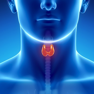
Shear-wave elastography (SWE) is an effective additional tool to complement conventional ultrasound for diagnosing Graves' disease, since it can characterize the tissue stiffness of the thyroid gland, according to a new study published in the May issue of the American Journal of Roentgenology.
The findings support the use of SWE to gather more information about thyroid tissue stiffness than may be possible with conventional ultrasound alone, wrote a team led by Dr. Shimei Li of Sun Yat-Sen University in Guangzhou, China.
"Conventional ultrasound can provide a variety of information, such as size, morphologic features, borders, internal echo characteristics, presence or absence of nodules, and blood flow conditions," the group wrote. "Recently, SWE has been proposed as a complementary technology to conventional ultrasound that can help provide tissue stiffness information. It not only provides a new reference index for the diagnosis of Graves' disease but also helps to monitor the changes in thyroid stiffness in different stages of disease progression."
Graves' disease tends to present in younger people and prompts overactivity of the thyroid gland, the team wrote. Ultrasound has long been used to diagnose the condition, revealing disease features such as an enlarged thyroid gland, diffuse limited hypoechogenicity, and intralesional vascularization on color Doppler (AJR, May 2019, Vol. 212:5, pp. 950-957).
But ultrasound elastography offers additional information about the thyroid gland's tissue stiffness, making SWE more objective than palpation, which is the typical manner of characterizing the tissue, the authors noted.
Li's group included 207 patients in the study. Of these, 162 had Graves' disease and 45 were healthy. All patients underwent an SWE exam that recorded three elasticity values in kilopascals (kPa) for each thyroid gland: SWE mean, SWE minimum, and SWE maximum.
The researchers found that the SWE elasticity values were higher in patients with Graves' disease than in healthy subjects.
| SWE elasticity values in patients with Graves' disease | ||
| Measure | Healthy subjects | Subjects with Graves' disease |
| SWE mean | 14.3 ± 2.7 kPa | 17.6 ± 6.4 kPa |
| SWE minimum | 8.4 ± 2.4 kPa | 10.7 ± 6.4 kPa |
| SWE maximum | 22.1 ± 5.4 kPa | 25.6 ± 10.6 kPa |
The team also found that the duration of disease, thyroid size, and isthmus thickness, as well as levels of thyroid peroxidase, thyroglobulin, and thyrotropin receptor antibodies correlated with SWE mean in patients with the disease.
It's possible that the results could help clinicians forecast how patients will respond to treatment, according to the group.
"Perhaps [assessing] the thyroid stiffness before treatment could predict the outcome of [radioactive iodine] treatment in patients with Graves' disease," the authors concluded.