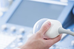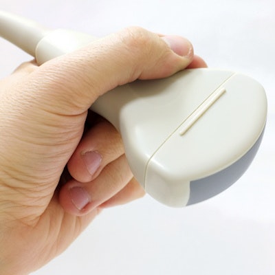
A short ergonomic training session can help sonographers learn to reduce their grip force on the ultrasound transducer, which could translate into fewer work-related musculoskeletal (MSK) injuries, according to a study published in the September/October issue of the Journal of Diagnostic Medical Sonography.
The findings suggest that a relatively simple intervention could help the up to 90% of sonographers who report work-related musculoskeletal pain, often due to how hard they grip the transducer, wrote a team led by Gill Harrison from City, University of London.
"Some sonographers working in specialized areas of practice, such as vascular or cardiac, are more likely to have wrist pain, which has been attributed to using a higher grip force," Harrison and colleagues wrote.
The researchers offered 13 sonographers a 30-minute ergonomic training session to explore whether this kind of intervention could reduce sonographers' grip force on the transducer. Each participant's maximum grip strength was initially measured using a dynamometer before he or she performed a transabdominal scan of a simulated case of early pregnancy. An ergometer was attached to the person's forearm, with the device making a sound when muscles were stimulated; the sound got louder as muscle strain increased. All participants scanned with their right hand (J Diagn Med Sonogr, September/October 2018, Vol. 34:5, pp. 321-327).
Harrison's group took photographs and video of each participant's hand, wrist, and arm while the person conducted the scan. Afterward, the team measured participants' grip force again by having them hold the dynamometer with the same force with which they had held the transducer.
The participants were then randomized into control (six sonographers) and intervention (seven sonographers) groups. Those in the intervention group reviewed the video or photographs of their hand and wrist to look for signs of tight grip; next, they performed the scan again and were measured again for grip force. Those in the control group did not review photographs or video of their hand and wrist during scanning, but they were also asked to scan again and were measured for grip force.
Twelve of the 13 study participants showed a statistically significant reduction in grip strength after the 30-minute ergonomic training session. However, the video/photo review did not result in further significant reduction in grip strength in the intervention group -- suggesting that the ergonomic session was more effective than a passive review of images.
"The results of this study demonstrated that ergonomic intervention had a significant impact on participants' matched grip force when holding the transducer after training," the group wrote.
A simple intervention could go a long way toward mitigating sonographers' injury risk, Harrison and colleagues concluded.
"Until new equipment developments are introduced to reduce the need for sonographers to use their own force to manipulate the transducer ... sonographers would benefit from some form of education to help them reduce their risk of injury," they wrote.