Dear AuntMinnie Member,
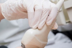
Ultrasound expert Dr. Jason Birnholz is back with his latest Practice of Ultrasound column. While the article focuses on imaging of the placenta, it also touches on one of the most important issues in healthcare: how much time physicians spend with patients.
Dr. Birnholz sees a disturbing subtext in medicine's focus on productivity and efficiency -- processes that often occur at the expense of the patient. He believes that attempts to shave minutes off ultrasound exams can impinge on efforts by ultrasound specialists to achieve the highest image quality, which can be critical in making a diagnosis.
Read Dr. Birnholz's thought-provoking comments by clicking here, or visit our Ultrasound Community at ultrasound.auntminnie.com.
Breast CT's bright future
While dedicated breast CT is still an investigational tool, researchers from New York and Massachusetts believe that it could develop into a modality well-suited for a wide variety of breast imaging tasks.
That's according to a new article in our Women's Imaging Community that explores the potential of this intriguing technology. Chief among breast CT's advantages is that it is a true 3D modality with better contrast and 3D anatomic detail, according to the researchers.
Concern about radiation dose has been one issue holding breast CT back, but in reality the radiation dose is comparable to that of combined exams with digital breast tomosynthesis and mammography, the group believes. Read more by clicking here.
Software-based FFR
In other news, be sure to visit our Advanced Visualization Community for an article on a new technique using a prototype software application for generating fractional flow reserve (FFR) images from CT data. Investigators believe it's a better alternative than two other techniques, one of which requires an invasive catheter to be placed near the heart, and another that requires data to be shipped to an offsite lab for processing. How well does it perform? Find out by clicking here, or visit the community at av.auntminnie.com.
Finally, we recently redesigned our website, and we hope you like its fresh and modern look. If you have any comments or feedback on the new design, please let us know in our Forums.