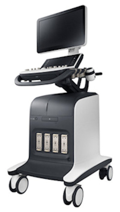
Samsung Electronics America has begun U.S. sales of its UGEO WS80A premium women's imaging ultrasound scanner.
The new UGEO WS80A ultrasound scanner. Image courtesy of Samsung.
The new system marks Samsung's entry into the high end of the women's imaging ultrasound segment, according to Doug Ryan, group vice president for health and medical equipment at Samsung's Enterprise Business Division. Until now, Samsung has offered scanners in the entry-level and midrange women's imaging segments, a legacy of its acquisition of Korean ultrasound developer Medison in 2010.
Key features of UGEO WS80A include what Samsung calls 5D imaging technology, an automated form of 4D (3D with motion) imaging. The technique enables users to create 5D images without much user interaction, addressing the issue of ultrasound being user-dependent. The technique is well-suited for applications such as myocardial perfusion index calculation, nuchal translucency imaging, and long-bone measurements to determine fetal age.
The 192-channel scanner also includes a 21.5-inch high-resolution LED monitor and better contrast and color visualization. Other highlights include Samsung's FRV (Feto Realistic View) technology for realistic depth perception of 3D anatomy, and single crystal probe design for better transducer penetration and resolution.
The system includes a five-year warranty, the first Samsung scanner with such extended protection, Ryan said. The company is launching the system at this week's American Institute of Ultrasound in Medicine (AIUM) meeting in Las Vegas.








![Examples of ultrasound findings and techniques. (A) Images in a 39-year-old male patient with a mass in the left thigh. The mass is heterogeneous on the B-mode US image (compared with the patient in D) and showed increased microvascularity (superb microvascular imaging [SMI]) and shear-wave elastography (SWE) values. Undifferentiated pleomorphic sarcoma was diagnosed at biopsy (with pleomorphic rhabdomyosarcoma in surgical specimen). (B) Images in an 18-year-old male patient with a mass in the left leg. The mass is hypoechoic on the B-mode image, with no other findings suggestive of malignancy. The lesion is in contact with the cortex of the tibia, which is slightly irregular. CT revealed a doubtful anteromedial tibial erosion. The microvascular study demonstrated high vascularization, suggestive of malignancy. Periosteal Ewing sarcoma was diagnosed with both histologic and immunohistochemical confirmation. (C) Images in a 69-year-old female patient with a lump growing on the outside of the left leg. Multiple SWE examinations were performed (please note the high values obtained in the measurements, whereas the color map highlights the stiffness relative to adjacent tissues). SMI showed areas of increased vascularization to target for sampling. Undifferentiated spindle cell sarcoma was diagnosed at biopsy, with residual leiomyosarcoma in the surgical specimen after neoadjuvant therapy. (D) Images in a 56-year-old female patient with a mass in the right thigh. The mass is heterogeneous at both B-mode ultrasound (similar to patient A) and MRI (coronal T2-weighted spectral attenuated inversion recovery [SPAIR]; T1-weighted pre-contrast and postcontrast imaging), which even shows uptake after the administration of paramagnetic contrast material, which is traditionally suggestive of malignancy. Low values at SMI and elastography are suggestive of benignity. Spindle cell lipoma was diagnosed at biopsy, with atypical spindle cell lipomatous tumor in the surgical specimen.](https://img.auntminnie.com/mindful/smg/workspaces/default/uploads/2026/08/images-radiol250278fig2.APCFLSvX6p.jpg?auto=format%2Ccompress&fit=crop&h=112&q=70&w=112)
