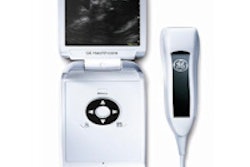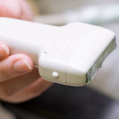
Broader training and thorough assessment of the benefits of point-of-care ultrasound are required before the technology can replace the stethoscope in physical examinations, according to an editorial published March 20 in the New England Journal of Medicine.
"Whether ultrasonography becomes as universally used and valued as [the stethoscope] will depend on whether training in its use becomes standard for future physicians and whether placing these devices in their hands is shown to improve medical practice at the point of care," wrote Dr. Scott Solomon and Dr. Fidencio Saldana of Brigham and Women's Hospital and Harvard Medical School.
The broadening use of compact ultrasound has served to "demystify and universalize ultrasonography," and a number of clinical studies have demonstrated that diagnostic ultrasound can outperform the traditional physical exam, according to the authors. In addition, several medical schools have begun offering ultrasound training early in the undergraduate curriculum (NEJM, March 20, 2014, Vol. 370:12, pp. 1083-1085).
Rigorous assessment of the benefits of point-of-care ultrasound and reporting of results will likely be required, however, for the next generation of physicians to widely incorporate ultrasound into the physical exam, according to Solomon and Saldana.
Furthermore, two major developments are needed before point-of-care ultrasound can replace the stethoscope, according to the authors. First, devices must become smaller, more ergonomic, and more functional, and second, a new generation of physicians must be trained in their use.
This won't be easy, as most new medical technologies tend to be reserved in medical school for subspecialists, who then train others in a top-down manner, according to the authors.
Furthermore, a number of practical issues will need to be considered, including whether ultrasound findings should be stored in a hospital's PACS and whether physicians should be allowed to bill for these procedures, they noted.
Of course, outfitting inexperienced practitioners with ultrasound is also controversial, owing to the increased risk of misdiagnosis. Substantial training is required to perform a competent ultrasound study, the authors wrote. For example, cardiology fellows typically need up to six weeks to learn the basic echocardiographic study.
Medical students may be able to make relatively crude diagnoses with ultrasound, but more sophisticated anatomical assessment will require substantially more training, they wrote. And the prospect of false-positive and false-negative findings raises issues.
There's also concern that ultrasound devices can distract students from the core principles of diagnosis and interpose another layer of technology between the doctor and patient, according to the authors.
They noted, though, that any new technology requiring training and expertise elicits "skepticism from practitioners steeped in older traditions."



![Examples of ultrasound findings and techniques. (A) Images in a 39-year-old male patient with a mass in the left thigh. The mass is heterogeneous on the B-mode US image (compared with the patient in D) and showed increased microvascularity (superb microvascular imaging [SMI]) and shear-wave elastography (SWE) values. Undifferentiated pleomorphic sarcoma was diagnosed at biopsy (with pleomorphic rhabdomyosarcoma in surgical specimen). (B) Images in an 18-year-old male patient with a mass in the left leg. The mass is hypoechoic on the B-mode image, with no other findings suggestive of malignancy. The lesion is in contact with the cortex of the tibia, which is slightly irregular. CT revealed a doubtful anteromedial tibial erosion. The microvascular study demonstrated high vascularization, suggestive of malignancy. Periosteal Ewing sarcoma was diagnosed with both histologic and immunohistochemical confirmation. (C) Images in a 69-year-old female patient with a lump growing on the outside of the left leg. Multiple SWE examinations were performed (please note the high values obtained in the measurements, whereas the color map highlights the stiffness relative to adjacent tissues). SMI showed areas of increased vascularization to target for sampling. Undifferentiated spindle cell sarcoma was diagnosed at biopsy, with residual leiomyosarcoma in the surgical specimen after neoadjuvant therapy. (D) Images in a 56-year-old female patient with a mass in the right thigh. The mass is heterogeneous at both B-mode ultrasound (similar to patient A) and MRI (coronal T2-weighted spectral attenuated inversion recovery [SPAIR]; T1-weighted pre-contrast and postcontrast imaging), which even shows uptake after the administration of paramagnetic contrast material, which is traditionally suggestive of malignancy. Low values at SMI and elastography are suggestive of benignity. Spindle cell lipoma was diagnosed at biopsy, with atypical spindle cell lipomatous tumor in the surgical specimen.](https://img.auntminnie.com/mindful/smg/workspaces/default/uploads/2026/08/images-radiol250278fig2.APCFLSvX6p.jpg?auto=format%2Ccompress&fit=crop&h=100&q=70&w=100)


![Examples of ultrasound findings and techniques. (A) Images in a 39-year-old male patient with a mass in the left thigh. The mass is heterogeneous on the B-mode US image (compared with the patient in D) and showed increased microvascularity (superb microvascular imaging [SMI]) and shear-wave elastography (SWE) values. Undifferentiated pleomorphic sarcoma was diagnosed at biopsy (with pleomorphic rhabdomyosarcoma in surgical specimen). (B) Images in an 18-year-old male patient with a mass in the left leg. The mass is hypoechoic on the B-mode image, with no other findings suggestive of malignancy. The lesion is in contact with the cortex of the tibia, which is slightly irregular. CT revealed a doubtful anteromedial tibial erosion. The microvascular study demonstrated high vascularization, suggestive of malignancy. Periosteal Ewing sarcoma was diagnosed with both histologic and immunohistochemical confirmation. (C) Images in a 69-year-old female patient with a lump growing on the outside of the left leg. Multiple SWE examinations were performed (please note the high values obtained in the measurements, whereas the color map highlights the stiffness relative to adjacent tissues). SMI showed areas of increased vascularization to target for sampling. Undifferentiated spindle cell sarcoma was diagnosed at biopsy, with residual leiomyosarcoma in the surgical specimen after neoadjuvant therapy. (D) Images in a 56-year-old female patient with a mass in the right thigh. The mass is heterogeneous at both B-mode ultrasound (similar to patient A) and MRI (coronal T2-weighted spectral attenuated inversion recovery [SPAIR]; T1-weighted pre-contrast and postcontrast imaging), which even shows uptake after the administration of paramagnetic contrast material, which is traditionally suggestive of malignancy. Low values at SMI and elastography are suggestive of benignity. Spindle cell lipoma was diagnosed at biopsy, with atypical spindle cell lipomatous tumor in the surgical specimen.](https://img.auntminnie.com/mindful/smg/workspaces/default/uploads/2026/08/images-radiol250278fig2.APCFLSvX6p.jpg?auto=format%2Ccompress&fit=crop&h=112&q=70&w=112)