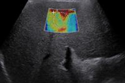
CHICAGO - High tumor stiffness at shear-wave elastography appears to predict lymph node metastasis in women with invasive breast cancer, and automated breast ultrasound (ABUS) turned in a similar diagnostic performance to handheld ultrasound, according to two studies presented on Sunday at the RSNA meeting.
Shear-wave elastography
Higher lesion stiffness values at shear-wave elastography have been shown to be linked with poorer prognoses, said Dr. Andrew Evans, of the University of Dundee in Scotland. Evans and colleagues conducted a study of 396 patients with invasive breast cancer who were treated by primary surgery and had shear-wave elastography at the time of diagnosis. Of this group, 217 were symptomatic, while 179 had screen-detected cancer.
The researchers used SuperSonic Imagine's Aixplorer ultrasound system to find mean stiffness values, taking measurements from a region of interest identified as the stiffest part of the abnormality. The team also assessed associations between lymph node involvement and mean lesion stiffness in kilopascals, invasive cancer size, histological grade, tumor type, estrogen (ER) and human epidermal growth factor (HER2) receptor status, and vascular invasion.
Median tumor size was 19 mm, and 28% of patients had lymph node involvement, Evans and colleagues found. On analysis, invasive size, histological grade, HER2 status, vascular invasion, tumor type, and mean stiffness were significantly associated with nodal involvement, Evans told session attendees.
"High tumor stiffness at shear-wave elastography is an independent predictor of lymph node involvement in women with invasive breast cancer," he said. "Our data may prove helpful for clinicians deciding which patients could benefit from neoadjuvant therapy."
ABUS matches handheld
During the same session, Dr. Eunjeong Kim, of the Catholic University of Korea, presented research indicating that automated breast ultrasound (ABUS) has similar diagnostic performance to handheld ultrasound.
Kim and colleagues evaluated 42 patients with 43 breast lesions who had been scheduled for an ultrasound-guided or stereotactic biopsy for suspicious calcifications on mammography. Each patient underwent both ABUS and handheld ultrasound.
Of the 43 lesions, 25 were malignant (58.1%) and 18 were benign (41.9%). There was no significant difference in the area under the curve (AUC) between automated breast ultrasound and handheld ultrasound (0.758 compared to 0.786, respectively).
"Automated breast ultrasound detected 96% of the malignant calcifications that had been found on mammography," Kim said. "Detection was found to be related to the pathology, calcification extent, and shape at mammography."


![Examples of ultrasound findings and techniques. (A) Images in a 39-year-old male patient with a mass in the left thigh. The mass is heterogeneous on the B-mode US image (compared with the patient in D) and showed increased microvascularity (superb microvascular imaging [SMI]) and shear-wave elastography (SWE) values. Undifferentiated pleomorphic sarcoma was diagnosed at biopsy (with pleomorphic rhabdomyosarcoma in surgical specimen). (B) Images in an 18-year-old male patient with a mass in the left leg. The mass is hypoechoic on the B-mode image, with no other findings suggestive of malignancy. The lesion is in contact with the cortex of the tibia, which is slightly irregular. CT revealed a doubtful anteromedial tibial erosion. The microvascular study demonstrated high vascularization, suggestive of malignancy. Periosteal Ewing sarcoma was diagnosed with both histologic and immunohistochemical confirmation. (C) Images in a 69-year-old female patient with a lump growing on the outside of the left leg. Multiple SWE examinations were performed (please note the high values obtained in the measurements, whereas the color map highlights the stiffness relative to adjacent tissues). SMI showed areas of increased vascularization to target for sampling. Undifferentiated spindle cell sarcoma was diagnosed at biopsy, with residual leiomyosarcoma in the surgical specimen after neoadjuvant therapy. (D) Images in a 56-year-old female patient with a mass in the right thigh. The mass is heterogeneous at both B-mode ultrasound (similar to patient A) and MRI (coronal T2-weighted spectral attenuated inversion recovery [SPAIR]; T1-weighted pre-contrast and postcontrast imaging), which even shows uptake after the administration of paramagnetic contrast material, which is traditionally suggestive of malignancy. Low values at SMI and elastography are suggestive of benignity. Spindle cell lipoma was diagnosed at biopsy, with atypical spindle cell lipomatous tumor in the surgical specimen.](https://img.auntminnie.com/mindful/smg/workspaces/default/uploads/2026/08/images-radiol250278fig2.APCFLSvX6p.jpg?auto=format%2Ccompress&fit=crop&h=100&q=70&w=100)


![Examples of ultrasound findings and techniques. (A) Images in a 39-year-old male patient with a mass in the left thigh. The mass is heterogeneous on the B-mode US image (compared with the patient in D) and showed increased microvascularity (superb microvascular imaging [SMI]) and shear-wave elastography (SWE) values. Undifferentiated pleomorphic sarcoma was diagnosed at biopsy (with pleomorphic rhabdomyosarcoma in surgical specimen). (B) Images in an 18-year-old male patient with a mass in the left leg. The mass is hypoechoic on the B-mode image, with no other findings suggestive of malignancy. The lesion is in contact with the cortex of the tibia, which is slightly irregular. CT revealed a doubtful anteromedial tibial erosion. The microvascular study demonstrated high vascularization, suggestive of malignancy. Periosteal Ewing sarcoma was diagnosed with both histologic and immunohistochemical confirmation. (C) Images in a 69-year-old female patient with a lump growing on the outside of the left leg. Multiple SWE examinations were performed (please note the high values obtained in the measurements, whereas the color map highlights the stiffness relative to adjacent tissues). SMI showed areas of increased vascularization to target for sampling. Undifferentiated spindle cell sarcoma was diagnosed at biopsy, with residual leiomyosarcoma in the surgical specimen after neoadjuvant therapy. (D) Images in a 56-year-old female patient with a mass in the right thigh. The mass is heterogeneous at both B-mode ultrasound (similar to patient A) and MRI (coronal T2-weighted spectral attenuated inversion recovery [SPAIR]; T1-weighted pre-contrast and postcontrast imaging), which even shows uptake after the administration of paramagnetic contrast material, which is traditionally suggestive of malignancy. Low values at SMI and elastography are suggestive of benignity. Spindle cell lipoma was diagnosed at biopsy, with atypical spindle cell lipomatous tumor in the surgical specimen.](https://img.auntminnie.com/mindful/smg/workspaces/default/uploads/2026/08/images-radiol250278fig2.APCFLSvX6p.jpg?auto=format%2Ccompress&fit=crop&h=112&q=70&w=112)