Contrast-enhanced ultrasound (CEUS) can effectively characterize solid focal liver lesions detected as incidental findings in the noncirrhotic liver, potentially avoiding the need for biopsy, according to research from the University of Trieste in Italy.
In a retrospective study of 55 patients, the group found that CEUS significantly improved diagnostic performance and reader confidence in characterizing solid focal liver lesions judged to be indeterminate after analysis of contrast-enhanced CT and/or MR. In 43 of 55 focal liver lesions, ultrasound findings would have obviated the need for biopsy.
"Contrast-enhanced ultrasound presents extremely high contrast resolution due to high sensitivity to the contrast [and] high spatial resolution due to the small field-of-view compared with CT and MR; you can concentrate exactly on the lesion," said Emilio Quaia, MD. "[It also provides] high temporal resolution, which allows the real-time assessment of contrast enhancement of the lesion."
Quaia presented the research during a scientific session at the 2010 RSNA meeting in Chicago.
Even though incidental focal liver lesions are mainly benign, they may present with equivocal enhancement patterns on contrast-enhanced CT or MR. This may occur because of incorrect scanning time or the true presence of atypical contrast enhancement or persistent lesion hypovascularity during the different dynamic phases, Quaia said.
"This frequently leads to low confidence in diagnosis and to biopsy or long-term follow-up," he said.
To assess whether contrast-enhanced ultrasound could effectively characterize these lesions, the research team retrospectively evaluated 55 incidental solid focal liver lesions in 25 noncirrhotic patients (20 female, five male; average age, 55 years; range, 35-87) over a five-year period. The lesions were 1-5 cm in diameter and were considered indeterminate after onsite analysis of multiphase contrast-enhanced CT and/or MR.
For inclusion in the study, each lesion had to present as equivocal or atypical enhancement during the arterial phase at contrast-enhanced CT/MR, or demonstrate hypovascularity on the portal late-phase or with persistent hypovascularity during all dynamic phases, Quaia said. In addition, contrast-enhanced ultrasound had to be performed within 25 days of the CT and/or MR exams.
Following injection of 2.4 mL of a commercially available ultrasound contrast agent (SonoVue, Bracco, Milan), each lesion was scanned by CEUS during the arterial (15-30 seconds after microbubble injection), portal-venous (40-75 seconds after injection), and late phase (80-180 seconds after injection).
Two blinded independent readers retrospectively reviewed the contrast-enhanced CT and/or MR studies before and after analysis of the contrast-enhanced ultrasound cine clips. They were asked to classify each lesion as malignant or benign according to standard diagnostic criteria based on the lesion's enhancement pattern at arterial phase and lesion vascularity on the portal and late phase, Quaia said.
 |
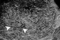
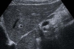
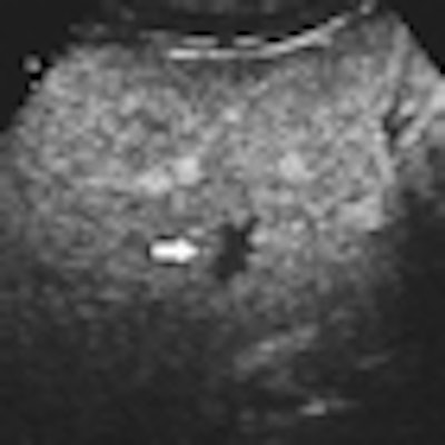
| Hemangioma with atypical enhancement on CT. (a) Contrast-enhanced CT, transverse plane. A solid lesion (arrow) appearing persistently hypovascular during the different dynamic phases (here represented during the portal-venous phase) is identified on the right liver. (b-d) Contrast-enhanced US after sulphur hexafluoride-filled microbubble injection during arterial (b), portal (c), and late phases (d). The lesion presents with globular peripheral enhancement during arterial phase (b), with a progressive centripetal fill-in during portal-venous (c) and late phase (d). Diagnosis of hemangioma was made after contrast-enhanced US. All images courtesy of Emilio Quaia, MD. |
 |
Final diagnoses for each lesion were available from imaging follow-up for 12 months in 48 lesions and histology in seven lesions. There were 40 hemangiomas, 12 metastases, two angiomyolipomas, and one hepatocellular adenoma.
Adding the review of contrast-enhanced ultrasound exams improved both the overall accuracy and confidence of the two radiologists in the study.
Accuracy for characterizing lesions
|
Diagnostic confidence (area under the ROC curve)
|
|||||||||
| ROC = receiver operator characteristics. The increases were statistically significant (p < 0.05). |
"Contrast-enhanced ultrasound improved the characterization of solid focal liver lesions appearing indeterminate on cross-sectional imaging by identifying some specific enhancement patterns, mainly nodular peripheral enhancement," Quaia said.
Peripheral nodular contrast enhancement during the arterial phase was noted in 25 lesions, and no contrast enhancement was seen in 18 lesions. Rimlike contrast enhancement was observed in eight lesions, and diffuse-central contrast enhancement was found in four lesions, Quaia said.
"But lesions appeared persistently hypovascular during the portal-venous and late phase," he said.
By Erik L. Ridley
AuntMinnie.com staff writer
January 28, 2011
Related Reading
US CAD boosts differentiation of focal liver lesions, December 17, 2010
Contrast US processing tool shows malignant liver lesions, July 30, 2010
Experience counts when reading contrast ultrasound liver images, January 19, 2010
CEUS aids differentiation of small liver lesions, April 3, 2006
Detection of liver metastases with contrast-enhanced ultrasound, March 23, 2006
Copyright © 2011 AuntMinnie.com