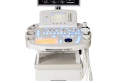
Philips Medical Systems of Andover, MA, will roll out several ultrasound systems and various new technologies in its product line, including enhancements to its flagship iU22 scanner and updates for the HD11 system.

Philips plans to update the HD11 platform by adding a flat-panel LCD monitor with articulating arm and adjustable keyboard for optimal ergonomic positioning, as well as cardiac capabilities that enable cardiologists to perform advanced quantification.
Also on display will be HD3, the smallest and most mobile of the company's HD ultrasound systems. It provides basic ultrasound users with more features, such as grayscale and color Doppler imaging with one-button image optimization, Cineloop review, tissue harmonic imaging, an adjustable flat-panel monitor, and analysis packages and measurements for various exams.
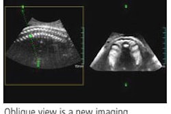
Among Philips' ancillary product highlights will be the X3-1 xMatrix array transducer for real-time 4D volume-rendered imaging for general and cardiac applications, the new S5-1 transducer, and 3D Viewer, which allows physicians to review, display, and rotate 3D datasets and create multiplanar reconstruction views similar to those used in MR and CT.
Other advanced analysis tools allow review of contrast loops and creation of microvascular imaging (MVI) files that visualize blood flow in extremely small vessels. Cardiac quantification of 3D datasets and strain quantification (SQ) are also available.
By Wayne Forrest
AuntMinnie.com contributing writer
November 9, 2005
Copyright © 2005 AuntMinnie.com








![Examples of ultrasound findings and techniques. (A) Images in a 39-year-old male patient with a mass in the left thigh. The mass is heterogeneous on the B-mode US image (compared with the patient in D) and showed increased microvascularity (superb microvascular imaging [SMI]) and shear-wave elastography (SWE) values. Undifferentiated pleomorphic sarcoma was diagnosed at biopsy (with pleomorphic rhabdomyosarcoma in surgical specimen). (B) Images in an 18-year-old male patient with a mass in the left leg. The mass is hypoechoic on the B-mode image, with no other findings suggestive of malignancy. The lesion is in contact with the cortex of the tibia, which is slightly irregular. CT revealed a doubtful anteromedial tibial erosion. The microvascular study demonstrated high vascularization, suggestive of malignancy. Periosteal Ewing sarcoma was diagnosed with both histologic and immunohistochemical confirmation. (C) Images in a 69-year-old female patient with a lump growing on the outside of the left leg. Multiple SWE examinations were performed (please note the high values obtained in the measurements, whereas the color map highlights the stiffness relative to adjacent tissues). SMI showed areas of increased vascularization to target for sampling. Undifferentiated spindle cell sarcoma was diagnosed at biopsy, with residual leiomyosarcoma in the surgical specimen after neoadjuvant therapy. (D) Images in a 56-year-old female patient with a mass in the right thigh. The mass is heterogeneous at both B-mode ultrasound (similar to patient A) and MRI (coronal T2-weighted spectral attenuated inversion recovery [SPAIR]; T1-weighted pre-contrast and postcontrast imaging), which even shows uptake after the administration of paramagnetic contrast material, which is traditionally suggestive of malignancy. Low values at SMI and elastography are suggestive of benignity. Spindle cell lipoma was diagnosed at biopsy, with atypical spindle cell lipomatous tumor in the surgical specimen.](https://img.auntminnie.com/mindful/smg/workspaces/default/uploads/2026/08/images-radiol250278fig2.APCFLSvX6p.jpg?auto=format%2Ccompress&dpr=2&fit=crop&h=167&q=70&w=250)