Dear AuntMinnie Member,
A work-in-progress ultrasound contrast agent has demonstrated good results in identifying sentinel lymph nodes. Researchers believe it offers a potential alternative to lymphoscintigraphy, a method commonly used to determine whether cancer has spread into the lymphatic system.
The news comes from a study presented this week at the Leading Edge in Diagnostic Ultrasound conference in Atlantic City, NJ, where staff writer Erik L. Ridley is reporting for our Ultrasound Digital Community.
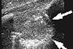
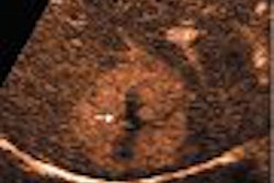
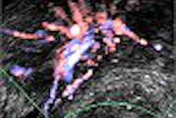
Researchers from Thomas Jefferson University in Philadelphia studied the contrast agent Sonazoid in an animal model to detect lymphatic drainage pathways in different anatomical regions. They found that normal sentinel lymph nodes demonstrated uniformly increased echogenicity, while SLNs with metastases showed nonuniform enhancement.
Lymphosonography also identified more SLNs than lymphoscintigraphy, although it fell short of the gold standard for SLN detection, blue-dye-guided surgical resection. Read all about the team's results by clicking here.
Another featured story from Leading Edge discusses the use of contrast-enhanced ultrasound in liver imaging, where Japanese researchers say it can significantly improve image quality to the point where ultrasound is competitive with CT and MRI. To view the article, visit our Ultrasound Digital Community at ultrasound.auntminnie.com.
![Examples of ultrasound findings and techniques. (A) Images in a 39-year-old male patient with a mass in the left thigh. The mass is heterogeneous on the B-mode US image (compared with the patient in D) and showed increased microvascularity (superb microvascular imaging [SMI]) and shear-wave elastography (SWE) values. Undifferentiated pleomorphic sarcoma was diagnosed at biopsy (with pleomorphic rhabdomyosarcoma in surgical specimen). (B) Images in an 18-year-old male patient with a mass in the left leg. The mass is hypoechoic on the B-mode image, with no other findings suggestive of malignancy. The lesion is in contact with the cortex of the tibia, which is slightly irregular. CT revealed a doubtful anteromedial tibial erosion. The microvascular study demonstrated high vascularization, suggestive of malignancy. Periosteal Ewing sarcoma was diagnosed with both histologic and immunohistochemical confirmation. (C) Images in a 69-year-old female patient with a lump growing on the outside of the left leg. Multiple SWE examinations were performed (please note the high values obtained in the measurements, whereas the color map highlights the stiffness relative to adjacent tissues). SMI showed areas of increased vascularization to target for sampling. Undifferentiated spindle cell sarcoma was diagnosed at biopsy, with residual leiomyosarcoma in the surgical specimen after neoadjuvant therapy. (D) Images in a 56-year-old female patient with a mass in the right thigh. The mass is heterogeneous at both B-mode ultrasound (similar to patient A) and MRI (coronal T2-weighted spectral attenuated inversion recovery [SPAIR]; T1-weighted pre-contrast and postcontrast imaging), which even shows uptake after the administration of paramagnetic contrast material, which is traditionally suggestive of malignancy. Low values at SMI and elastography are suggestive of benignity. Spindle cell lipoma was diagnosed at biopsy, with atypical spindle cell lipomatous tumor in the surgical specimen.](https://img.auntminnie.com/mindful/smg/workspaces/default/uploads/2026/08/images-radiol250278fig2.APCFLSvX6p.jpg?auto=format%2Ccompress&fit=crop&h=100&q=70&w=100)


![Examples of ultrasound findings and techniques. (A) Images in a 39-year-old male patient with a mass in the left thigh. The mass is heterogeneous on the B-mode US image (compared with the patient in D) and showed increased microvascularity (superb microvascular imaging [SMI]) and shear-wave elastography (SWE) values. Undifferentiated pleomorphic sarcoma was diagnosed at biopsy (with pleomorphic rhabdomyosarcoma in surgical specimen). (B) Images in an 18-year-old male patient with a mass in the left leg. The mass is hypoechoic on the B-mode image, with no other findings suggestive of malignancy. The lesion is in contact with the cortex of the tibia, which is slightly irregular. CT revealed a doubtful anteromedial tibial erosion. The microvascular study demonstrated high vascularization, suggestive of malignancy. Periosteal Ewing sarcoma was diagnosed with both histologic and immunohistochemical confirmation. (C) Images in a 69-year-old female patient with a lump growing on the outside of the left leg. Multiple SWE examinations were performed (please note the high values obtained in the measurements, whereas the color map highlights the stiffness relative to adjacent tissues). SMI showed areas of increased vascularization to target for sampling. Undifferentiated spindle cell sarcoma was diagnosed at biopsy, with residual leiomyosarcoma in the surgical specimen after neoadjuvant therapy. (D) Images in a 56-year-old female patient with a mass in the right thigh. The mass is heterogeneous at both B-mode ultrasound (similar to patient A) and MRI (coronal T2-weighted spectral attenuated inversion recovery [SPAIR]; T1-weighted pre-contrast and postcontrast imaging), which even shows uptake after the administration of paramagnetic contrast material, which is traditionally suggestive of malignancy. Low values at SMI and elastography are suggestive of benignity. Spindle cell lipoma was diagnosed at biopsy, with atypical spindle cell lipomatous tumor in the surgical specimen.](https://img.auntminnie.com/mindful/smg/workspaces/default/uploads/2026/08/images-radiol250278fig2.APCFLSvX6p.jpg?auto=format%2Ccompress&fit=crop&h=112&q=70&w=112)