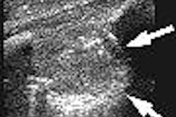
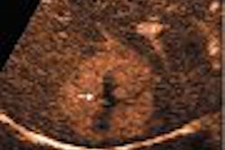
Ultrasound of the abdomen can help diagnose dengue fever, and in the event of a dengue epidemic, the ultrasound features of thickened gallbladder wall, pleural effusion, and ascites should strongly favor diagnosis of the condition, according to researchers from India.
Dengue is endemic in more than 100 countries and threatens the health of 40% of the world's population. India has seen 50 outbreaks of the epidemic so far, according to the researchers from the Sri Ramachandra Medical College & Research Institute in Chennai.
"Ultrasound of the abdomen is an important adjunct to clinical profile in diagnosing DF (dengue fever) and may help to direct further confirmatory investigations," the researchers wrote in their study published in the British Journal of Radiology (May 2005, Vol. 78:929, pp. 416-418).
The retrospective study included 128 patients suspected of dengue fever, 88 of whom tested serologically positive. The researchers used a Symphony portable ultrasound system (L&T Medical, Mumbai, India) with 3.5-MHz and 5-MHz probes.
They found thickened gallbladder with pericholecystic fluid to be the most common initial ultrasound finding in the infected patients.
The distinguishing ultrasound features noted on scans performed up to the seventh day of the onset of fever in the 88 serologically positive patients were thickened gallbladder in all patients, followed by right pleural effusion in 71.9% of the patients, ascites in 53.2%, left pleural effusion in 21.9%, and pericardial effusion 28.5%. Hepatomegaly was also observed in 34.3% and splenomegaly in 15.6% of the serologically positive patients.
In one group of 32 serologically positive patients, ultrasound was performed between the second and third days after onset of fever, and followed up with scans between the fifth and seventh days. In the second group of 56 serologically positive patients, ultrasound was performed only between the fifth and seventh days.
In the first group, seven patients had hepatomegaly (21.8%), two had splenomegaly (6.25%), and two had right pleural effusion (6.25%). All patients had gallbladder wall thickening and pericholecystic fluid, and none had ascites, left pleural effusion, or pericardial effusion.
On the follow-up scan performed between the fifth and seventh days, however, ascites were observed in 17 patients (53.1%), left-sided pleural effusion in seven patients (21.8%), and pericardial effusion in nine patients (28.1%). Hepatomegaly was observed in four more patients (12.5%), splenomegaly in three more patients (9.37%), and right-sided pleural effusion in 21 more patients (65.6%). Gallbladder wall thickening persisted on the follow-up scans.
In the second group, the researchers found hepatomegaly in 34 patients (60.9%), splenomegaly in 17 (30.4%), ascites in four (7.14%), right pleural effusion in 55 (96.5%), left pleural effusion in 37 (66.1%), and pericardial effusion in 16 (28.6%). Gallbladder thickening was present in all patients.
The clinical and laboratory findings confirmed diagnosis of classical dengue fever in 65 patients, dengue hemorrhagic fever in 19 patients, and dengue shock syndrome in four. Among the 40 patients who were serologically negative, the gallbladder pathology observed in the initial ultrasound scans of 14 patients disappeared on the follow-up scans, the researchers noted.
Advantages and limitations
Serology is the mainstay in diagnosing dengue fever, but diagnosis is often delayed due to the time needed to get the results. With ultrasound, further diagnosis can be made early in the course of the disease compared with other modes of diagnosis, the researchers stated.
The major limitation, however, is that gallbladder thickening is a nonspecific finding when considered in isolation and occurs in other viral infections, enteric fever, and leptospirosis as well. "But in other viral infections, the historical profile, symptom complex evolution, and physical findings do not mimic those of DF," the researchers wrote.
In enteric fever, they noted, ultrasound features include splenomegaly, intra-abdominal lymphadenopathy, intramural thickening of the terminal ileum and cecum, and renal abnormalities such as arteriectasis and perinephric fluid collection, in addition to gallbladder wall thickening and polyserositis. In leptospirosis, gross abnormalities involving hepatic and renal parenchyma are observed.
"During an epidemic the ultrasound findings of GB (gallbladder) wall thickening with or without polyserositis in a febrile patient should suggest the possibility of DF/DHF (dengue hemorrhagic fever)," the researchers concluded.
By N. Shivapriya
AuntMinnie.com staff writer
May 17, 2005
Related Reading
Ultrasound helpful in dengue fever diagnosis, December 18, 2003
3D ultrasound offers reliable assessment of gallbladder, bile ducts, May 5, 2003
Preoperative assessment of bile duct stones with sonography, November 7, 2002
Ultrasound differentiates causes of abdominal pain in kids, October 4, 2002
Ultrasound separates galactoceles from simple cysts, April 9, 2002
Copyright © 2005 AuntMinnie.com