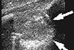
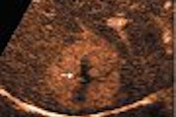
ATLANTIC CITY - Contrast-enhanced ultrasound is showing promise for identifying sentinel lymph nodes (SLNs), and may offer accuracy improvements over lymphoscintigraphy, according to a presentation Tuesday at the Leading Edge in Diagnostic Ultrasound conference.
"(Our) experiments clearly show that lymphosonography could be utilized in a variety of animal models to detect lymphatic drainage pathways in numerous anatomical regions," according to Dr. Barry Goldberg, a professor of radiology at Thomas Jefferson University (TJU) in Philadelphia.
Following up on previous TJU research that examined the use of contrast-enhanced ultrasound to detect SLNs in swine with naturally occurring melanoma tumors, a TJU study team sought to determine if lymphosonography could be applied after subcutaneous, submucosal, or parenchymal injections of the same contrast agent in various anatomical locations in other animal species.
Twenty animals were used in the experiments, which utilized Sonazoid (GE Healthcare, Chalfont St. Giles, U.K.) as well as an ultrasound scanner with a 7.5-MHz flat linear-array transducer for transcutaneous imaging and a 6.5-MHz endocavity transducer for endorectal imaging. The researchers performed fundamental 2D grayscale and color-flow imaging, as well as grayscale phase-inversion harmonic imaging and multiplanar 3D ultrasound scans.
Normal SLNs demonstrated uniformly increased echogenicity, Goldberg said. SLNs with metastases, however, demonstrated nonuniform enhancement. The researchers were able to identify SLNs as small as 4 mm.
"Using lymphosonography, we can detect SLNs and lymphatic channels in several different animal species," he said. "Enhancement of SLNs was seen after subcutaneous, submucosal, and intraparenchymal injections. With lymph node sonography, dissection is not required to follow lymphatic channels to their location."
The researchers were also able to answer a previously unanswered question on what was the mechanism of uptake and retention of contrast microbubbles within the lymphatic system. Using scanning electron microscopy, the study team injected the hind footpads of two rabbits with Sonazoid.
Ultrastructural analysis of the contrast-enhanced SLNs revealed multiple intracytoplasmic spherical vacuoles within the macrophages, Goldberg said. This was not seen within the control group.
The vacuoles ranged in size from 1.07 ųm to 1.99 ųm, with variations relating to different planes of section through the spherical structures.
TJU has received a three-year grant from the National Institutes of Health to study lymphosonography, an effort that began January 1 and will include more than 124 animals.
To date under the grant-related research, 12 swine with 28 melanomas have been evaluated for the project with ultrasound, lymphoscintigraphy, and blue-dye injections. Lymphosonography identified 55 SLNs, with one false-positive finding.
Lymphoscintigraphy identified 38 hot spots suspected of representing SLNs, including two false-positive and two second-echelon nodes. Blue-dye-guided surgical resection identified 62 lymph nodes, two of which were determined to represent second-echelon nodes, Goldberg said.
Ultrasound detected SLNs ranging in size from 3-32 mm. Of the 26 SLNs not seen with lymphoscintigraphy, 21 were detected with lymphosonography, he said. Only one SLN detected with lymphoscintigraphy was not identified by lymphosonography.
Overall, lymphosonography accurately detected SLNs in 90% of cases, compared with a 57% rate for lymphoscintigraphy.
Future TJU research will use the melanoma swine model to identify the minimum amount of tumor in SLNs that can be identified by lymphosonography, and the average number of SLNs per tumor, Goldberg said.
By Erik L. Ridley
AuntMinnie.com staff writer
May 12, 2005
Related Reading
Intraoperative imprint cytology advised for suspicious axillary lymph nodes, March 10, 2005
U.K. to introduce sentinel node biopsy, October 27, 2004
Nomogram predicts breast cancer spread beyond sentinel lymph node, January 8, 2004
Detection of occult metastases in head and neck cancer feasible with SLN biopsy, October 20, 2003
Copyright © 2005 AuntMinnie.com

![Examples of ultrasound findings and techniques. (A) Images in a 39-year-old male patient with a mass in the left thigh. The mass is heterogeneous on the B-mode US image (compared with the patient in D) and showed increased microvascularity (superb microvascular imaging [SMI]) and shear-wave elastography (SWE) values. Undifferentiated pleomorphic sarcoma was diagnosed at biopsy (with pleomorphic rhabdomyosarcoma in surgical specimen). (B) Images in an 18-year-old male patient with a mass in the left leg. The mass is hypoechoic on the B-mode image, with no other findings suggestive of malignancy. The lesion is in contact with the cortex of the tibia, which is slightly irregular. CT revealed a doubtful anteromedial tibial erosion. The microvascular study demonstrated high vascularization, suggestive of malignancy. Periosteal Ewing sarcoma was diagnosed with both histologic and immunohistochemical confirmation. (C) Images in a 69-year-old female patient with a lump growing on the outside of the left leg. Multiple SWE examinations were performed (please note the high values obtained in the measurements, whereas the color map highlights the stiffness relative to adjacent tissues). SMI showed areas of increased vascularization to target for sampling. Undifferentiated spindle cell sarcoma was diagnosed at biopsy, with residual leiomyosarcoma in the surgical specimen after neoadjuvant therapy. (D) Images in a 56-year-old female patient with a mass in the right thigh. The mass is heterogeneous at both B-mode ultrasound (similar to patient A) and MRI (coronal T2-weighted spectral attenuated inversion recovery [SPAIR]; T1-weighted pre-contrast and postcontrast imaging), which even shows uptake after the administration of paramagnetic contrast material, which is traditionally suggestive of malignancy. Low values at SMI and elastography are suggestive of benignity. Spindle cell lipoma was diagnosed at biopsy, with atypical spindle cell lipomatous tumor in the surgical specimen.](https://img.auntminnie.com/mindful/smg/workspaces/default/uploads/2026/08/images-radiol250278fig2.APCFLSvX6p.jpg?auto=format%2Ccompress&fit=crop&h=100&q=70&w=100)


![Examples of ultrasound findings and techniques. (A) Images in a 39-year-old male patient with a mass in the left thigh. The mass is heterogeneous on the B-mode US image (compared with the patient in D) and showed increased microvascularity (superb microvascular imaging [SMI]) and shear-wave elastography (SWE) values. Undifferentiated pleomorphic sarcoma was diagnosed at biopsy (with pleomorphic rhabdomyosarcoma in surgical specimen). (B) Images in an 18-year-old male patient with a mass in the left leg. The mass is hypoechoic on the B-mode image, with no other findings suggestive of malignancy. The lesion is in contact with the cortex of the tibia, which is slightly irregular. CT revealed a doubtful anteromedial tibial erosion. The microvascular study demonstrated high vascularization, suggestive of malignancy. Periosteal Ewing sarcoma was diagnosed with both histologic and immunohistochemical confirmation. (C) Images in a 69-year-old female patient with a lump growing on the outside of the left leg. Multiple SWE examinations were performed (please note the high values obtained in the measurements, whereas the color map highlights the stiffness relative to adjacent tissues). SMI showed areas of increased vascularization to target for sampling. Undifferentiated spindle cell sarcoma was diagnosed at biopsy, with residual leiomyosarcoma in the surgical specimen after neoadjuvant therapy. (D) Images in a 56-year-old female patient with a mass in the right thigh. The mass is heterogeneous at both B-mode ultrasound (similar to patient A) and MRI (coronal T2-weighted spectral attenuated inversion recovery [SPAIR]; T1-weighted pre-contrast and postcontrast imaging), which even shows uptake after the administration of paramagnetic contrast material, which is traditionally suggestive of malignancy. Low values at SMI and elastography are suggestive of benignity. Spindle cell lipoma was diagnosed at biopsy, with atypical spindle cell lipomatous tumor in the surgical specimen.](https://img.auntminnie.com/mindful/smg/workspaces/default/uploads/2026/08/images-radiol250278fig2.APCFLSvX6p.jpg?auto=format%2Ccompress&fit=crop&h=112&q=70&w=112)