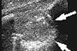
(Radiology Review) Sonographic measurement and appearance of the testicular appendage can be used to diagnose torsion of the testicular appendage, according to Brazilian researchers.
Dr. Matteo Baldisserotto and colleagues at Hospital da Criança Conceição in Porto Alegre determined an upper limit of normal measurement for the testicular appendage, as well as normal and abnormal grayscale and Doppler appearances in a study recently published in the American Journal of Roentgenology.
Differentiating between torsed testis and testicular appendage is an important clinical challenge because of the high morbidity associated with torsion of the testis. Defining the size of normal and torsed testicular appendages with ultrasound imaging is a useful and safe diagnostic tool for determining torsion of testicular appendages, enabling conservative management and reduction in false-positive results for testicular torsion, the researchers stated.
Thirty-three male children with acute scrotal pain were preoperatively examined using ultrasound imaging. Torsion of scrotal contents was found in 27 patients and six had epididymitis. Twenty-two patients had torsion of the appendix testis, three had torsion of the testis, and two had torsion of the appendix epididymis.
Using grayscale and color Doppler, the team demonstrated the testicular appendage in 23 patients. Most of these patients had a torsed testicular appendage. "With the visualization of an appendix larger than 5.6 mm as the sonographic diagnostic criterion for torsion of a testicular appendage, sensitivity was 68.2%, and specificity was 100%," the authors wrote. There were two patients with epididymitis and enlarged appendix testis (16.0 mm and 9.2 mm), and one torsed appendix testis measuring 5 mm, which was not demonstrated on ultrasound.
The researchers described the sonographic appearances in 21 patients with torsed appendix testis as hyperechoic to the epididymis in 11 patients, hypoechoic in three, heterogenous in four, and isoechoic in three. "The two normal testicular appendages found in the patients with epididymitis were hypoechoic," they stated.
"The identification of a testicular appendage larger than 5.6 mm is suggestive of torsion. Therefore, depending on the patients' clinical conditions, these cases can be treated conservatively when an appendage larger than 5.6 mm is identified," the researchers concluded.
"Color Doppler Sonography of Normal and Torsed Testicular Appendages in Children"
Baldisserotto, Matteo, et al
Hospital da Criança Conceição, Ministério da Saúde - Brazil, Eca de Queiroz, 384 Apt. 502, Porto Alegre, RS, Brazil, 90.670-020.
AJR 2005 (April); 184:1287-1292
By Radiology Review
April 21, 2005
Copyright © 2005 AuntMinnie.com



![Examples of ultrasound findings and techniques. (A) Images in a 39-year-old male patient with a mass in the left thigh. The mass is heterogeneous on the B-mode US image (compared with the patient in D) and showed increased microvascularity (superb microvascular imaging [SMI]) and shear-wave elastography (SWE) values. Undifferentiated pleomorphic sarcoma was diagnosed at biopsy (with pleomorphic rhabdomyosarcoma in surgical specimen). (B) Images in an 18-year-old male patient with a mass in the left leg. The mass is hypoechoic on the B-mode image, with no other findings suggestive of malignancy. The lesion is in contact with the cortex of the tibia, which is slightly irregular. CT revealed a doubtful anteromedial tibial erosion. The microvascular study demonstrated high vascularization, suggestive of malignancy. Periosteal Ewing sarcoma was diagnosed with both histologic and immunohistochemical confirmation. (C) Images in a 69-year-old female patient with a lump growing on the outside of the left leg. Multiple SWE examinations were performed (please note the high values obtained in the measurements, whereas the color map highlights the stiffness relative to adjacent tissues). SMI showed areas of increased vascularization to target for sampling. Undifferentiated spindle cell sarcoma was diagnosed at biopsy, with residual leiomyosarcoma in the surgical specimen after neoadjuvant therapy. (D) Images in a 56-year-old female patient with a mass in the right thigh. The mass is heterogeneous at both B-mode ultrasound (similar to patient A) and MRI (coronal T2-weighted spectral attenuated inversion recovery [SPAIR]; T1-weighted pre-contrast and postcontrast imaging), which even shows uptake after the administration of paramagnetic contrast material, which is traditionally suggestive of malignancy. Low values at SMI and elastography are suggestive of benignity. Spindle cell lipoma was diagnosed at biopsy, with atypical spindle cell lipomatous tumor in the surgical specimen.](https://img.auntminnie.com/mindful/smg/workspaces/default/uploads/2026/08/images-radiol250278fig2.APCFLSvX6p.jpg?auto=format%2Ccompress&fit=crop&h=100&q=70&w=100)


![Examples of ultrasound findings and techniques. (A) Images in a 39-year-old male patient with a mass in the left thigh. The mass is heterogeneous on the B-mode US image (compared with the patient in D) and showed increased microvascularity (superb microvascular imaging [SMI]) and shear-wave elastography (SWE) values. Undifferentiated pleomorphic sarcoma was diagnosed at biopsy (with pleomorphic rhabdomyosarcoma in surgical specimen). (B) Images in an 18-year-old male patient with a mass in the left leg. The mass is hypoechoic on the B-mode image, with no other findings suggestive of malignancy. The lesion is in contact with the cortex of the tibia, which is slightly irregular. CT revealed a doubtful anteromedial tibial erosion. The microvascular study demonstrated high vascularization, suggestive of malignancy. Periosteal Ewing sarcoma was diagnosed with both histologic and immunohistochemical confirmation. (C) Images in a 69-year-old female patient with a lump growing on the outside of the left leg. Multiple SWE examinations were performed (please note the high values obtained in the measurements, whereas the color map highlights the stiffness relative to adjacent tissues). SMI showed areas of increased vascularization to target for sampling. Undifferentiated spindle cell sarcoma was diagnosed at biopsy, with residual leiomyosarcoma in the surgical specimen after neoadjuvant therapy. (D) Images in a 56-year-old female patient with a mass in the right thigh. The mass is heterogeneous at both B-mode ultrasound (similar to patient A) and MRI (coronal T2-weighted spectral attenuated inversion recovery [SPAIR]; T1-weighted pre-contrast and postcontrast imaging), which even shows uptake after the administration of paramagnetic contrast material, which is traditionally suggestive of malignancy. Low values at SMI and elastography are suggestive of benignity. Spindle cell lipoma was diagnosed at biopsy, with atypical spindle cell lipomatous tumor in the surgical specimen.](https://img.auntminnie.com/mindful/smg/workspaces/default/uploads/2026/08/images-radiol250278fig2.APCFLSvX6p.jpg?auto=format%2Ccompress&fit=crop&h=112&q=70&w=112)