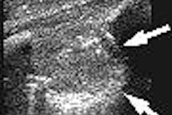
(Radiology Review) Benign prostatic hyperplasia originating in the outer gland may mimic prostate cancer, according to researchers in China. Both pathologies appear on endorectal ultrasound as hypoechoic, so transrectal ultrasound-guided biopsy should be used to provide histological differentiation, according to Dr. Jie Tang and colleagues. Their study appeared in the April edition of the Journal of Ultrasound in Medicine.
Previous research and traditional thinking has dictated that benign hyperplastic areas only appeared in the central prostate gland, the authors stated. During a 42-month period, they performed prostate biopsies in 472 patients suspected of having prostate cancer. Prior to endorectal sonography, patients were given a cleansing enema and antibiotic prophylaxis. Sonography was performed using a 6- to 10-MHz endorectal transducer with the patient in the left lateral decubitus position.
The team at the Chinese People's Liberation Army General Hospital in Beijing determined that while the majority of the 240 patients with hypoechoic nodules had prostate cancer, 22 patients (9.17%) had biopsy-proven focal benign hyperplasia within the outer gland.
Patients without hypoechoic nodules had sextant biopsies performed, while patients with nodules had two biopsies per site followed by sextant biopsies. Tissue samples were taken using an automated core biopsy device, a Biopty gun (C.R. Bard, Covington, GA), with an 18-gauge needle. "Focal nodules were seen as well-circumscribed with an ovoid shape and smooth surface in 18 patients," they stated. Among all the patients, 310 (65.7%) had a benign histologic finding, and 162 (34.32%) had a histologic diagnosis of prostate cancer. The majority of benign hypoechoic nodules on ultrasound were smooth, well-circumscribed oval lesions with an average diameter of 1.4 cm.
Anatomically, Dr. Jie Tang and colleagues described the prostatic outer gland as a combination of the central zone and peripheral zone, as the two zones are not easily distinguished sonographically, even using high-resolution endorectal transducers.
"Benign hyperplasia may sometimes originate in the prostatic outer gland, and may appear as a hypoechoic nodule, similar to the appearance of prostate cancer," the authors concluded. They recommended further analysis of the histologic differences comparing hyperplasia in the inner gland with hyperplasia in the outer gland.
"Correlation between Hypoechoic Nodules on Ultrasonography and Benign Hyperplasia in the Prostatic Outer Gland"
Tang, Jie, et al
Department of Ultrasound, Chinese People's Liberation Army General Hospital, 28 Fuxing Road, Beijing 100853, China
J Ultrasound Med 2005 (April) 24:483-488
By Radiology Review
April 15, 2005
Copyright © 2005 AuntMinnie.com



![Examples of ultrasound findings and techniques. (A) Images in a 39-year-old male patient with a mass in the left thigh. The mass is heterogeneous on the B-mode US image (compared with the patient in D) and showed increased microvascularity (superb microvascular imaging [SMI]) and shear-wave elastography (SWE) values. Undifferentiated pleomorphic sarcoma was diagnosed at biopsy (with pleomorphic rhabdomyosarcoma in surgical specimen). (B) Images in an 18-year-old male patient with a mass in the left leg. The mass is hypoechoic on the B-mode image, with no other findings suggestive of malignancy. The lesion is in contact with the cortex of the tibia, which is slightly irregular. CT revealed a doubtful anteromedial tibial erosion. The microvascular study demonstrated high vascularization, suggestive of malignancy. Periosteal Ewing sarcoma was diagnosed with both histologic and immunohistochemical confirmation. (C) Images in a 69-year-old female patient with a lump growing on the outside of the left leg. Multiple SWE examinations were performed (please note the high values obtained in the measurements, whereas the color map highlights the stiffness relative to adjacent tissues). SMI showed areas of increased vascularization to target for sampling. Undifferentiated spindle cell sarcoma was diagnosed at biopsy, with residual leiomyosarcoma in the surgical specimen after neoadjuvant therapy. (D) Images in a 56-year-old female patient with a mass in the right thigh. The mass is heterogeneous at both B-mode ultrasound (similar to patient A) and MRI (coronal T2-weighted spectral attenuated inversion recovery [SPAIR]; T1-weighted pre-contrast and postcontrast imaging), which even shows uptake after the administration of paramagnetic contrast material, which is traditionally suggestive of malignancy. Low values at SMI and elastography are suggestive of benignity. Spindle cell lipoma was diagnosed at biopsy, with atypical spindle cell lipomatous tumor in the surgical specimen.](https://img.auntminnie.com/mindful/smg/workspaces/default/uploads/2026/08/images-radiol250278fig2.APCFLSvX6p.jpg?auto=format%2Ccompress&fit=crop&h=100&q=70&w=100)


![Examples of ultrasound findings and techniques. (A) Images in a 39-year-old male patient with a mass in the left thigh. The mass is heterogeneous on the B-mode US image (compared with the patient in D) and showed increased microvascularity (superb microvascular imaging [SMI]) and shear-wave elastography (SWE) values. Undifferentiated pleomorphic sarcoma was diagnosed at biopsy (with pleomorphic rhabdomyosarcoma in surgical specimen). (B) Images in an 18-year-old male patient with a mass in the left leg. The mass is hypoechoic on the B-mode image, with no other findings suggestive of malignancy. The lesion is in contact with the cortex of the tibia, which is slightly irregular. CT revealed a doubtful anteromedial tibial erosion. The microvascular study demonstrated high vascularization, suggestive of malignancy. Periosteal Ewing sarcoma was diagnosed with both histologic and immunohistochemical confirmation. (C) Images in a 69-year-old female patient with a lump growing on the outside of the left leg. Multiple SWE examinations were performed (please note the high values obtained in the measurements, whereas the color map highlights the stiffness relative to adjacent tissues). SMI showed areas of increased vascularization to target for sampling. Undifferentiated spindle cell sarcoma was diagnosed at biopsy, with residual leiomyosarcoma in the surgical specimen after neoadjuvant therapy. (D) Images in a 56-year-old female patient with a mass in the right thigh. The mass is heterogeneous at both B-mode ultrasound (similar to patient A) and MRI (coronal T2-weighted spectral attenuated inversion recovery [SPAIR]; T1-weighted pre-contrast and postcontrast imaging), which even shows uptake after the administration of paramagnetic contrast material, which is traditionally suggestive of malignancy. Low values at SMI and elastography are suggestive of benignity. Spindle cell lipoma was diagnosed at biopsy, with atypical spindle cell lipomatous tumor in the surgical specimen.](https://img.auntminnie.com/mindful/smg/workspaces/default/uploads/2026/08/images-radiol250278fig2.APCFLSvX6p.jpg?auto=format%2Ccompress&fit=crop&h=112&q=70&w=112)