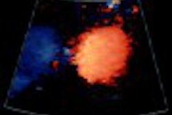
(Ultrasound Review) In a recent American Journal of Roentgenology study of soft-tissue tumors in infancy, high-frequency grayscale and color Doppler imaging were used to evaluate distinguishing features.
"Birthmarks of various shades of red, blue, of pink represent vascular tumors or malformations, and a distinction between them is based on the detection of cellular proliferation in tumors and its absence in malformations," reported radiologists and dermatologists at the University of Montreal in Quebec, Canada. Technical advancements in image quality and vascular sensitivity have enabled sonography to be used to evaluate the subcutaneous portion of these lesions more accurately.
The group studied 16 children with vascular tumors, excluding hemangiomas, which were scheduled for biopsy diagnosis. The ultrasound characteristics evaluated included margin definition, echogenicity, number of blood vessels, resistive index, and peak systolic velocity. The ultrasound characteristics were compared with the biopsy findings.
There were five hemangioendotheliomas, six tufted angiomas, and five infantile myofibromatoses. Tufted angiomas were echogenic, while infantile myofibromatoses were isoechoic or hypoechoic. None of the tufted angiomas or infantile myofibromatoses had calcifications, but four of five hemangioendotheliomas had calcifications. Hemangioendotheliomas and tufted angiomas were ill-defined lesions, while in contrast infantile myofibromatoses that were well defined, they reported.
Comparing vascularity, tufted angiomas and infantile myofibromatoses were the least vascularized of the three tumor types, with low velocity flow and low vessel density. In most cases of hemangioendotheliomas, the vessel density was estimated as medium and the peak systolic frequency shift was greater than 2k Hz. A previous study by the same authors demonstrated that hemangiomas could be differentiated from other masses by evaluating vascular characteristics with ultrasound. Hemangiomas have a high vessel density (>five vessels/cm2), and peak systolic Doppler shift greater than 2k Hz, they found.
The authors concluded that "the vascular tumors -- hemangioendotheliomas, tufted angiomas, and infantile myofibromatoses -- were distinguishable from hemangiomas on Doppler sonography in all cases except one hemangioendothelioma. Unlike hemangiomas, these lesions should be investigated by biopsy or excision."
Vascular soft-tissue tumors in infancy: distinguishing features on Doppler sonographyJ Dubois et al
Department of medical imaging, Hôpital Sainte-Justine and University of Montreal, Montreal, Quebec, Canada
AJR 2002 June; 178:1541-1545
By Ultrasound Review
July 24, 2002
Copyright © 2002 AuntMinnie.com

![Examples of ultrasound findings and techniques. (A) Images in a 39-year-old male patient with a mass in the left thigh. The mass is heterogeneous on the B-mode US image (compared with the patient in D) and showed increased microvascularity (superb microvascular imaging [SMI]) and shear-wave elastography (SWE) values. Undifferentiated pleomorphic sarcoma was diagnosed at biopsy (with pleomorphic rhabdomyosarcoma in surgical specimen). (B) Images in an 18-year-old male patient with a mass in the left leg. The mass is hypoechoic on the B-mode image, with no other findings suggestive of malignancy. The lesion is in contact with the cortex of the tibia, which is slightly irregular. CT revealed a doubtful anteromedial tibial erosion. The microvascular study demonstrated high vascularization, suggestive of malignancy. Periosteal Ewing sarcoma was diagnosed with both histologic and immunohistochemical confirmation. (C) Images in a 69-year-old female patient with a lump growing on the outside of the left leg. Multiple SWE examinations were performed (please note the high values obtained in the measurements, whereas the color map highlights the stiffness relative to adjacent tissues). SMI showed areas of increased vascularization to target for sampling. Undifferentiated spindle cell sarcoma was diagnosed at biopsy, with residual leiomyosarcoma in the surgical specimen after neoadjuvant therapy. (D) Images in a 56-year-old female patient with a mass in the right thigh. The mass is heterogeneous at both B-mode ultrasound (similar to patient A) and MRI (coronal T2-weighted spectral attenuated inversion recovery [SPAIR]; T1-weighted pre-contrast and postcontrast imaging), which even shows uptake after the administration of paramagnetic contrast material, which is traditionally suggestive of malignancy. Low values at SMI and elastography are suggestive of benignity. Spindle cell lipoma was diagnosed at biopsy, with atypical spindle cell lipomatous tumor in the surgical specimen.](https://img.auntminnie.com/mindful/smg/workspaces/default/uploads/2026/08/images-radiol250278fig2.APCFLSvX6p.jpg?auto=format%2Ccompress&fit=crop&h=100&q=70&w=100)


![Examples of ultrasound findings and techniques. (A) Images in a 39-year-old male patient with a mass in the left thigh. The mass is heterogeneous on the B-mode US image (compared with the patient in D) and showed increased microvascularity (superb microvascular imaging [SMI]) and shear-wave elastography (SWE) values. Undifferentiated pleomorphic sarcoma was diagnosed at biopsy (with pleomorphic rhabdomyosarcoma in surgical specimen). (B) Images in an 18-year-old male patient with a mass in the left leg. The mass is hypoechoic on the B-mode image, with no other findings suggestive of malignancy. The lesion is in contact with the cortex of the tibia, which is slightly irregular. CT revealed a doubtful anteromedial tibial erosion. The microvascular study demonstrated high vascularization, suggestive of malignancy. Periosteal Ewing sarcoma was diagnosed with both histologic and immunohistochemical confirmation. (C) Images in a 69-year-old female patient with a lump growing on the outside of the left leg. Multiple SWE examinations were performed (please note the high values obtained in the measurements, whereas the color map highlights the stiffness relative to adjacent tissues). SMI showed areas of increased vascularization to target for sampling. Undifferentiated spindle cell sarcoma was diagnosed at biopsy, with residual leiomyosarcoma in the surgical specimen after neoadjuvant therapy. (D) Images in a 56-year-old female patient with a mass in the right thigh. The mass is heterogeneous at both B-mode ultrasound (similar to patient A) and MRI (coronal T2-weighted spectral attenuated inversion recovery [SPAIR]; T1-weighted pre-contrast and postcontrast imaging), which even shows uptake after the administration of paramagnetic contrast material, which is traditionally suggestive of malignancy. Low values at SMI and elastography are suggestive of benignity. Spindle cell lipoma was diagnosed at biopsy, with atypical spindle cell lipomatous tumor in the surgical specimen.](https://img.auntminnie.com/mindful/smg/workspaces/default/uploads/2026/08/images-radiol250278fig2.APCFLSvX6p.jpg?auto=format%2Ccompress&fit=crop&h=112&q=70&w=112)