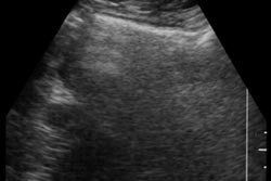
(Ultrasound Review) - This prospective study of 23 neonates, infants and young children with lower abdominal cystic masses assessed the value of the 'daughter cyst' sign in diagnosing ovarian cysts.
The researchers defined the 'daughter cyst' sign as the presence of a small cyst along the wall of a cystic mass. In neonates, infants and young children, normal ovaries have small cystic masses that represent unstimulated follicles, but the authors sought to show that the daughter cyst sign would distinguish the follicles of ovarian cysts.
Other pelvic cystic masses include: intestinal duplication cyst, meconium pseudocyst, hydrometrocolpos, lymphangioma, anterior meningeocele, mesenteric cyst and urachal cyst. These are mostly congenital cysts requiring surgical removal. Ovarian cysts, in contrast, can spontaneously resolve during the first few months of life. Surgical intervention may be avoided if the correct diagnosis of ovarian cyst is made.
Ultrasound examinations were performed using a 5-MHz curved array or 7-MHz linear array transducer and an Acuson 128XP. The walls of the cysts were carefully imaged in search of daughter cysts. These small cysts can either protrude into the lumen of the large cystic mass or lie along the outer wall.
Daughter cysts were demonstrated in 82% of the patients with ovarian cysts. Seven involuted during the first seven months of life, and in the remaining four girls, the cysts were surgically removed. Pathology confirmed they were ovarian follicles.
Results indicated that the daughter cyst sign showed a sensitivity of 82% and a specificity of 100% in differentiating ovarian cysts from other pelvic cysts.
"The daughter cyst sign is a specific sonographic finding for an ovarian cyst and may be useful in differentiating uncomplicated ovarian cysts from other cystic masses in neonates, infants, and young children," the authors conclude.
" 'Daughter cyst' sign: a sonographic finding of ovarian cyst in neonates, infants, and young children," by Hee-Jung Lee et al, Dept of Diagnostic Radiology, Keimyung Universty School of Medicine, Dongsan Medical Center, 194, Dongsan-dong, Jung-Ku,Taegu 700-310, Korea. AJR 2000; 174:1013-1015.
By Ultrasound Review
August 16, 2000

![Examples of ultrasound findings and techniques. (A) Images in a 39-year-old male patient with a mass in the left thigh. The mass is heterogeneous on the B-mode US image (compared with the patient in D) and showed increased microvascularity (superb microvascular imaging [SMI]) and shear-wave elastography (SWE) values. Undifferentiated pleomorphic sarcoma was diagnosed at biopsy (with pleomorphic rhabdomyosarcoma in surgical specimen). (B) Images in an 18-year-old male patient with a mass in the left leg. The mass is hypoechoic on the B-mode image, with no other findings suggestive of malignancy. The lesion is in contact with the cortex of the tibia, which is slightly irregular. CT revealed a doubtful anteromedial tibial erosion. The microvascular study demonstrated high vascularization, suggestive of malignancy. Periosteal Ewing sarcoma was diagnosed with both histologic and immunohistochemical confirmation. (C) Images in a 69-year-old female patient with a lump growing on the outside of the left leg. Multiple SWE examinations were performed (please note the high values obtained in the measurements, whereas the color map highlights the stiffness relative to adjacent tissues). SMI showed areas of increased vascularization to target for sampling. Undifferentiated spindle cell sarcoma was diagnosed at biopsy, with residual leiomyosarcoma in the surgical specimen after neoadjuvant therapy. (D) Images in a 56-year-old female patient with a mass in the right thigh. The mass is heterogeneous at both B-mode ultrasound (similar to patient A) and MRI (coronal T2-weighted spectral attenuated inversion recovery [SPAIR]; T1-weighted pre-contrast and postcontrast imaging), which even shows uptake after the administration of paramagnetic contrast material, which is traditionally suggestive of malignancy. Low values at SMI and elastography are suggestive of benignity. Spindle cell lipoma was diagnosed at biopsy, with atypical spindle cell lipomatous tumor in the surgical specimen.](https://img.auntminnie.com/mindful/smg/workspaces/default/uploads/2026/08/images-radiol250278fig2.APCFLSvX6p.jpg?auto=format%2Ccompress&fit=crop&h=100&q=70&w=100)


![Examples of ultrasound findings and techniques. (A) Images in a 39-year-old male patient with a mass in the left thigh. The mass is heterogeneous on the B-mode US image (compared with the patient in D) and showed increased microvascularity (superb microvascular imaging [SMI]) and shear-wave elastography (SWE) values. Undifferentiated pleomorphic sarcoma was diagnosed at biopsy (with pleomorphic rhabdomyosarcoma in surgical specimen). (B) Images in an 18-year-old male patient with a mass in the left leg. The mass is hypoechoic on the B-mode image, with no other findings suggestive of malignancy. The lesion is in contact with the cortex of the tibia, which is slightly irregular. CT revealed a doubtful anteromedial tibial erosion. The microvascular study demonstrated high vascularization, suggestive of malignancy. Periosteal Ewing sarcoma was diagnosed with both histologic and immunohistochemical confirmation. (C) Images in a 69-year-old female patient with a lump growing on the outside of the left leg. Multiple SWE examinations were performed (please note the high values obtained in the measurements, whereas the color map highlights the stiffness relative to adjacent tissues). SMI showed areas of increased vascularization to target for sampling. Undifferentiated spindle cell sarcoma was diagnosed at biopsy, with residual leiomyosarcoma in the surgical specimen after neoadjuvant therapy. (D) Images in a 56-year-old female patient with a mass in the right thigh. The mass is heterogeneous at both B-mode ultrasound (similar to patient A) and MRI (coronal T2-weighted spectral attenuated inversion recovery [SPAIR]; T1-weighted pre-contrast and postcontrast imaging), which even shows uptake after the administration of paramagnetic contrast material, which is traditionally suggestive of malignancy. Low values at SMI and elastography are suggestive of benignity. Spindle cell lipoma was diagnosed at biopsy, with atypical spindle cell lipomatous tumor in the surgical specimen.](https://img.auntminnie.com/mindful/smg/workspaces/default/uploads/2026/08/images-radiol250278fig2.APCFLSvX6p.jpg?auto=format%2Ccompress&fit=crop&h=112&q=70&w=112)