
A study of over 2,100 older adults found that specific brain MRI features, particularly tightness of the high-convexity sulci, are significantly associated with cognitive decline over five to seven years, suggesting these imaging markers could help identify people at risk for memory loss before symptoms develop.
- Baseline tightness of high-convexity sulci (THC) on brain MRI showed significant associations with cognitive decline in older adults aged 65 and over.
- Study included 2,133 community-based participants followed for 5-7 years with baseline MRI scans and cognitive assessments.
- 46.3% of participants showed incomplete DESH features while only 1.7% had complete DESH characteristics.
- THC demonstrated nearly 3 times higher odds of cognitive decline (OR 2.95) in cognitively normal older adults.
- Researchers recommend careful MRI assessment of THC for early risk stratification and prevention of cognitive impairment in aging populations.
Certain brain MRI features may signal cognitive decline, suggest findings published October 13 in Radiology.
More specifically, baseline tightness of the high-convexity sulci (THC) at MRI is tied to cognitive decline in adults aged 65 years or older, wrote researchers led by Kana Saito, MD, from Hirosaki University in Japan.
“These findings suggest that careful assessment of THC may aid early risk stratification,” Saito and colleagues wrote.
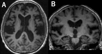
Disproportionately enlarged subarachnoid space hydrocephalus (DESH) is a neuroimaging characteristic of idiopathic normal pressure hydrocephalus (iNPH), a progressive neurologic condition that can lead to cognitive impairment. DESH includes three key morphologic components: ventricular enlargement (Evans index greater than 0.3), THC, and enlargement of the Sylvian fissures.
Prior research suggests associations between complete DESH and Mini-Mental State Examination score decline. And automated DESH scoring algorithms have shown negative correlations between these scores and cognitive function in older populations.
However, many individuals do not show all three key DESH features and may only show one or two features, known as incomplete DESH.
The Saito team studied the prevalence of complete and incomplete DESH and their respective associations with baseline cognitive impairment and follow-up cognitive decline.
 Images in a 75-year-old female participant with complete disproportionately enlarged subarachnoid space hydrocephalus. (A) Axial and (B) coronal T1-weighted MRI scans demonstrate enlargement of the Sylvian fissures, tightness of the high-convexity sulci, and an Evans index greater than 0.3 (Evans index = 0.31).RSNA
The study included a community-based cohort of 2,133 participants aged 65 years and older. The participants underwent baseline brain MRI and were followed for five to seven years. Two radiologists assessed baseline MRI scans for core DESH features.
Of the total participants, 37 had complete DESH (1.7%), 988 had incomplete DESH (46.3%), and 1,108 had no DESH (51.9%).
The researchers found no evidence linking baseline imaging features to baseline cognitive impairment (all Holm-adjusted p > 0.05).
However, baseline THC showed significant associations with follow-up cognitive decline in the cognitively normal cohort (odds ratio [OR], 2.95; Holm-adjusted p < 0.001) and in a combined cohort participants with normal cognition or mild cognitive impairment (OR, 2.87; Holm-adjusted p < 0.001).
Finally, THC plus enlarged Sylvian fissures revealed associations with decline only in the combined cohort (OR, 1.83; Holm-adjusted p = 0.04).
The study authors called for future studies to incorporate longitudinal imaging. This approach could “clarify the mechanisms underlying THC” and show THC’s role in early risk stratification for cognitive decline.
The results extend findings on how imaging features may be tied to cognitive decline, according to an accompanying editorial written by Sven Haller, MD, from the Cornavin Medical Imaging Center in Geneva, Switzerland.
“I expect that the coexistence of imaging features of incomplete normal pressure hydrocephalus configuration, glymphatic system anomaly, neurodegenerative disease, and cerebrovascular disease is common, and that these conditions interact in a nonlinear and supra-additive way,” Haller wrote. “This may accelerate the risk of cognitive de-cline and potentially confound research studies and clinical trials that focus, for example, on isolated Alzheimer’s disease pathology, ignoring existing co-pathology.”
Read the full study here.

.fFmgij6Hin.png?auto=compress%2Cformat&fit=crop&h=100&q=70&w=100)


.fFmgij6Hin.png?auto=compress%2Cformat&fit=crop&h=167&q=70&w=250)


