An AI-enhanced single-shot cine MRI algorithm for assessing left ventricular function in patients with irregular heartbeat produces image quality and ventricular measurements comparable to those produced by conventional cine MRI, researchers have reported.
The findings could improve imaging results in this population, wrote a team led by Nan Zhang, a supervisor radiologic technologist at Zhongshan Hospital of Fudan University in Shanghai, China. The study was published March 26 in Radiology: Cardiothoracic Imaging.
"For patients with severe arrhythmias, breath-holding can be particularly challenging, often resulting in compromised image quality, exam failure, or inaccurate assessments," Zhang said in an RSNA statement.
The accurate evaluation of left ventricular function in patients with heart failure is key to determining treatment decisions, monitoring therapeutic responses, and predicting outcomes, the team explained. Cardiac MRI is the go-to technique for this task and is traditionally performed using conventional cine sequences called balanced steady-state free precession -- an approach that captures moving images and visualizes bodily processes in real time but requires patients to hold their breath multiple times during the scan, which can be difficult for those with arrhythmia, or irregular heartbeat.
The group evaluated the feasibility of a shorter technique to assess individuals with arrhythmia called retrospective electrocardiographically (ECG) gated single-shot cine using deep learning-enhanced compressed sensing (AI-CS). They compared it to conventional balanced steady-state free precession (bSSFP) cine. The research included 70 participants who underwent short-axis cine imaging with both bSSFP and AI-CS single-shot sequences on a 1.5-tesla MRI scanner between September 2023 and September 2024. Of these 70 individuals, 25 were healthy volunteers and 45 had suspected arrhythmia.
The researchers measured left ventricular volumetric and strain parameters measured by both methods; parameters included end-diastolic volume, end-systolic volume, stroke volume, ejection fraction, peak strain (in radial, longitudinal, and circumferential directions), and standard deviation of peak strain. Three cardiovascular radiologists analyzed the images, assessing artifacts and the visibility of cardiac structures. These readers were blinded to clinical information and prior imaging results.
Overall, the team found that the success rate for the AI-CS single-shot cine sequence was 100%, compared to 88% for the conventional cine sequence. Furthermore, the AI-CS technique reduced scan time from 132 seconds to 10 seconds.
The group also reported the following:
- The AI-CS single-shot cine showed better image quality compared to conventional cine, particularly in participants with arrhythmia, with fewer "mistrigger" events (when the scanner misses an intended phase due to the patient's irregular heartbeat) and motion artifacts.
- AI-CS also demonstrated good-to-excellent agreement with conventional cine for measurements of biventricular volumes and left ventricular mass.
- In cases where conventional cine failed, AI-CS provided ejection fraction -- the measurement of how much blood the heart's left ventricular pumps out -- comparable to values from echocardiography.
"The AI-CS sequence effectively avoided the cardiac motion artifacts commonly caused by mistriggering in conventional cine," Zhang said. "It also demonstrated a shorter mean acquisition time while providing improved image quality, particularly in the visualization of the endocardial border, epicardial border, papillary muscles, and cardiac motion."
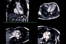
 Representative images of participants with difficulty with breath holding and arrhythmia. In the lower right corner of A–L, the position coordinates of the images are marked. Short-axis (SA) view images in the end-systole (A–F) and end-diastole (G–L) phases in participants with atrial fibrillation. (A–C, G–I) Balanced steady-state free precession (bSSFP) cardiac cine. (D–F, J–L) Deep learning–enhanced compressed sensing (AI-CS) single-shot cardiac cine. In contrast to AI-CS single-shot cardiac cine, endocardial contours were challenging to identify on conventional segmented cine in midventricular and apical sections. (M) Line graph of time curve during the whole cardiac cycle obtained from bSSFP cardiac cine. (N) Line graph of time curve during the whole cardiac cycle obtained from AI-CS single-shot cardiac cine. LV = left ventricle; 2D = two-dimensional.RSNA
The takeaway? The AI-CS framework offers a promising alternative for cardiac MRI examinations in the clinical setting, where long acquisition time remains a major challenge, according to Zhang.
"Further optimization of the AI-CS framework, particularly regarding image contrast and artifact reduction, will enhance its applicability in routine clinical practice," he concluded.
Access the full study here.