An ultrafast MRI imaging method appears capable of high-resolution, multicontrast imaging and parametric mapping of the entire human brain in less than three minutes, according to a project presented May 14 at the International Society for Magnetic Resonance in Medicine (ISMRM) 2025 meeting.
A team at the Biomedical Imaging Research Institute at Cedars-Sinai Medical Center in Los Angeles developed the framework that synthesized high-resolution, whole-brain multicontrast MR images, along with T1 and T2 maps from a combination of low-resolution images and a single high-resolution image.
The approach could improve and facilitate diagnosis and monitoring of neurological diseases, such as multiple sclerosis (MS), by making detailed brain imaging feasible in time-sensitive clinical settings, according to the team.
In testing, statistical and visual results indicated good performance, however with room for improvement, noted Beril Alyuz, who presented the group's magna cum laude merit award-winning work.
While multicontrast MRI and quantitative (q) MRI have been developed to speed up MR imaging of the brain, each has its shortcomings, Alyuz explained. In addition, specialized sequences and complex reconstruction algorithms are usually not available on standard MRI scanners, she said.
Toward an alternative, the group developed a unified deep-learning framework designed for both contrast generation and qMRI map estimation using significantly accelerated scans. Existing methods typically focus on one or the other, Alyuz said.
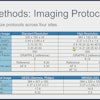
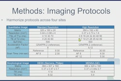
Input acquisition protocol consisted of high-resolution T1-weighted imaging for detailed spatial information, in addition to low-resolution T2-weighted, T2-star weighted, and diffusion-weighted imaging (DWI), as part of a U-Net architecture network nearly identical to one used in a previous study, according to Alyuz.
Against a reference acquisition protocol of 11 qualitative images routinely used for brain MRI and two quantitative maps, the input acquisition protocol took less than 3 minutes compared to the reference protocol's 53 minutes, Alyuz said.
Preprocessing involved bias correction, image registration, skull stripping, and normalization, along with data augmentation with modality dropout. Modality dropout helps with overfitting, provides robustness for missing contrasts at the input, and guides the network to use the higher resolution input in the absence of the others, Alyuz explained.
Described as a multibranch, unit-based network with a deeper path for high-resolution T1-weighted images and separate decoder heads for separate output channels, the network is designed to be adaptive, according to Alyuz.
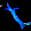
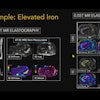
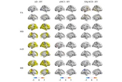
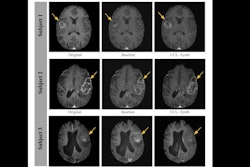
The team used their method to reconstruct the (Magnetom, Siemens Healthineers) scans of 15 healthy volunteers and three people with MS. Alyuz reported satisfactory image quality and accuracy in both groups, adding that the multicontrast MR images demonstrated successful reconstruction of pathological details related to MS.
Contrast weighted images were evaluated with [structural similarity index] SSIM, [peak signal to noise ratio] PSNR, and [normalized root mean squared error] NRMSE. For T1 and T2 maps, [masked auto encoders] MAE, NRMSE, SSIM, and PSNR were calculated across the full brain.
 Beril Alyuz, ISMRM 2025
"The only clear degradation was in the white matter, which might indicate that our model was sensitive to the lesions, but this might be explained by the lack of MS patients in the training data," Alyuz said.
 Beril Alyuz, ISMRM 2025
The model's generalizability could be enhanced with a larger dataset including a variety of neurological disorders, she added. Further, clinical validation with a larger patient population is needed, and the team plans to conduct a clinical trial toward this end.
Principal investigators of the project are Yibin Xie, PhD, and Biomedical Imaging Research Institute Director Debiao Li, PhD.
"Current fast imaging approaches typically require specialized sequences and/or reconstructions that are limited to certain vendors, scanners, and software versions," Li told AuntMinnie. "And usually, they do not include quantitative imaging such as T1/T2 mappings, which have shown added diagnostic value for many diseases.
"We are developing a different approach, based on deep learning, to effectively combine widely available sequences to generate high-resolution, multi-contrast, and quantitative brain MRI," Li continued. "It is designed to be generalizable, without requiring highly specialized, often exclusive sequences. The end product is an ultrafast (2.5 mins) brain MRI scan that can be used for a general brain assessment on most platforms."
Check out AuntMinnie’s full coverage of ISMRM 2025 here.