Researchers have used MRI to illuminate the effects of hypertension on cerebral small vessel disease-related brain structures, finding that these effects differ by patient sex and age at diagnosis.
The results could help tailor patient care, wrote a team led by doctoral candidate Amanpreet Kaur of McGill University Health Centre in Montreal, Canada. The group's results were published on December 19 in Hypertension.
"Our findings demonstrate the significance of how age at hypertension diagnosis affects cerebrovascular health differentially in males and females via small vessel disease … [and could] help identify sex-specific neuroimaging biomarkers that can inform clinical practice and public health guidelines in the management of hypertension and brain structural changes, which could lead to cognitive decline later in life," the team noted.
Previous research has shown that an individual's age at hypertension diagnosis can contribute to structural brain changes associated with cerebral small vessel disease, it wrote.
"Younger age at onset of hypertension, independent of blood pressure control, is predictive of cognitive decline and increased vascular dementia risk in later life," the group explained. "Moreover, high blood pressure in early adulthood and into midlife is associated with late life reductions in brain volume and white matter hyperintensities, as measured by MRI, both of which are hallmarks of cerebral small vessel disease."
Yet whether high blood pressure's effects on cognitive health differ by sex has been unclear, according to and colleagues. To address the knowledge gap, they evaluated sex differences and any associations between age at hypertension diagnosis and cerebral small vessel disease-related brain structural changes.
Their study included data from 9,430 patients in the UK Biobank who had a known age of hypertension diagnosis and had undergone brain MR imaging. The study participants were categorized by sex and age at hypertension diagnosis. The group also included data from a matched control cohort of patients who had brain MR imaging but did not have hypertension. The investigators tracked morphological brain structural changes and assessed the white matter microstructure of the two groups.
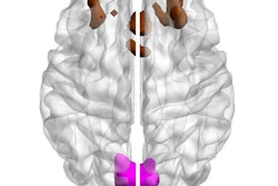
The researchers discovered that men who had been diagnosed with high blood pressure at a younger age had lower brain gray and white matter volume compared to individuals who had not, and that the volume of white matter hyperintensities was greater in both men and women with hypertension than in men and women without it.
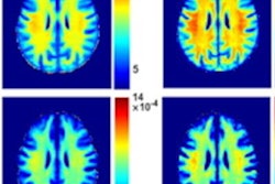
"Compared with normotensive controls, white matter microstructural integrity was lower in individuals with hypertension, which became more prominent with increasing age," the team reported.
| White matter microstructural integrity as measured by factor analysis, or FA (principal component analysis outcome factors 1 and 2) in hypertensive participants as compared to normotensive participants. |
|||
|---|---|---|---|
| FA_PCA Factors (mean, sd) | Hypertensive | Normotensive | P-value |
| PC Factor 1 (highest variance) | -0.09 | 0.09 | <0.0001 |
| PC Factor 2 (second highest variance) | -0.008 | 0.008 | 0.3 |
"Midlife hypertension is a significant, modifiable factor affecting brain volume," the group wrote.
More research is needed, according to Kaur's team.
"[Sex]-specific analyses are required to determine the effect of hypertension on cerebral small vessel disease-related brain changes in males and females," it concluded.