A machine-learning model used with breast MR images shows that higher levels of background parenchymal enhancement (BPE) point to increased breast cancer risk in women with extremely dense breasts, a study published August 8 in Radiology has found.
The findings offer another way to assess breast cancer risk in women with dense tissue, wrote a team led by Hui Wang, MD, of the University Medical Center Utrecht in the Netherlands.
"Automated identification of quantitative breast parenchymal enhancement features on dynamic contrast-enhanced (DCE) MRI scans could provide added value for assessing breast cancer risk in women with extremely dense breasts," the group wrote.
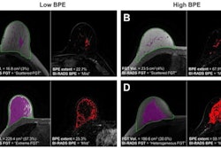
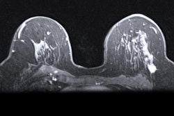
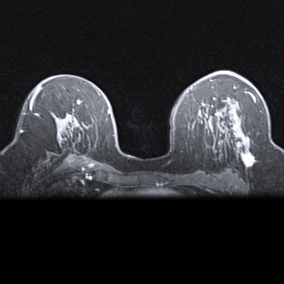
Breast MRI is used as an additional screening tool in women with dense tissue, as mammography can miss early-stage breast cancer in this population, the team noted. But in addition to density, another breast cancer risk factor is BPE – that is, the level of normal fibroglandular tissue that enhances on breast MRI, Wang and colleagues wrote. Connections between BPE and breast cancer risk have been underexplored.
"Thus far, studies on breast cancer risk factors have typically focused on women at high lifetime risk of developing breast cancer," study co-author Kenneth Gilhuijs, PhD, of the University Medical Center Utrecht in the Netherlands said in a statement released by the RSNA. "This is the first study that we know of that demonstrates an association between background parenchymal enhancement and occurrence of breast cancer in women with extremely dense breasts."
Wang and colleagues conducted a study that included data from 4,553 women who participated in the Dense Tissue and Early Breast Neoplasm Screening (DENSE) trial, a study that sought to develop a deep-learning model that would identify BPE. The women underwent contrast-enhanced MRI exams every two years in eight hospitals in the Netherlands between December 2011 and January 2016. The researchers adjusted for age, body mass index, and BPE.
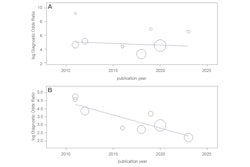
The key finding was that the combination of the artificial intelligence algorithm and MR imaging showed that the incidence of breast cancer was greater in women with higher volumes of BPE compared with women with lower volumes. Of the 4,553 women who participated in the study, 122 received a breast cancer diagnosis, and 63% of those were diagnosed after the first round of screening, the group reported.
Machine learning could help tailor screening for women with dense breast tissue, Gilhuijs told AuntMinnie.com.
"If the results of our study reproduce in other populations, machine learning of breast parenchymal enhancement may become a useful objective tool in clinical practice to help radiologists improve the assessment of a woman's individual risk of breast cancer in concert with other known risk factors," he said. "This could ultimately lead ... to tailoring the screening frequency of women more precisely to their individual risk."
The study fills in a knowledge gap in the literature, wrote Lars Grimm, MD, of Duke University in Durham, NC, in an accompanying commentary.
"This study fits nicely into the growing body of evidence of breast parenchyma as an important predictor of breast cancer risk," Grimm noted. "[The research] demonstrates the value of breast parenchymal measurements and shows that important information can be extracted from the images, beyond subjective radiologist assessment of BPE."
The complete study can be found here.