
Portable MRI developer Hyperfine is collaborating with the International Society for Magnetic Resonance in Medicine (ISMRM) and the Bill & Melinda Gates Foundation on an educational program to increase neuroimaging clinical research and knowledge transfer between high-income and low- and medium-income countries.
A goal of the ISMRM program is to facilitate local neuroimaging capabilities in low- and medium-income countries by enabling access to ISMRM's educational programs, and by establishing a global mentorship program for its clinicians.
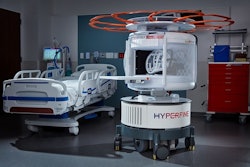
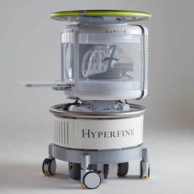
Hyperfine explains in a press release that this also includes the deployment of MR brain imaging equipment made possible by an existing partnership with the Bill & Melinda Gates Foundation's UNITY Project. This Gates Foundation funded-initiative allows leading researchers to study environmental factors affecting early brain development, and it is focused on neurodevelopment patterns in sub-Saharan and South Africa. As part of that project, Hyperfine has already deployed its Swoop portable MRI scanners in Bangladesh, Ethiopia, Ghana, Kenya, Malawi, Pakistan, South Africa, and Uganda.
As part of the program, the ISMRM-Gates Foundation partnership underwrote the attendance of 100 participants who were members of the UNITY Project and/or the newly formed Africa chapter of the ISMRM to attend ISMRM 2023. They have also been paired with individual long-term mentors, which include three Hyperfine managers: Chief Medical Officer and Chief Strategy Officer Khan Siddiqui, MD; Senior Research Scientist Megan Poorman, PhD; and Francesco Padormo, PhD.

![Overview of the study design. (A) The fully automated deep learning framework was developed to estimate body composition (BC) (defined as subcutaneous adipose tissue [SAT] in liters; visceral adipose tissue [VAT] in liters; skeletal muscle [SM] in liters; SM fat fraction [SMFF] as a percentage; and intramuscular adipose tissue [IMAT] in deciliters) from MRI. The fully automated framework comprised one model (model 1) to quantify different BC measures (SAT, VAT, SM, SMFF, and IMAT) as three-dimensional (3D) measures from whole-body MRI scans. The second model (model 2) was trained to identify standardized anatomic landmarks along the craniocaudal body axis (z coordinate field), which allowed for subdividing the whole-body measures into different subregions typically examined on clinical routine MRI scans (chest, abdomen, and pelvis). (B) BC was quantified from whole-body MRI in over 66,000 individuals from two large population-based cohort studies, the UK Biobank (UKB) (36,317 individuals) and the German National Cohort (NAKO) (30,291 individuals). Bar graphs show age distribution by sex and cohort. BMI = body mass index. (C) After the performance assessment of the fully automated framework, the change in BC measures, distributions, and profiles across age decades were investigated. Age-, sex-, and height-adjusted body composition reference curves were calculated and made publicly available in a web-based z-score calculator (https://circ-ml.github.io).](https://img.auntminnie.com/mindful/smg/workspaces/default/uploads/2026/05/body-comp.XgAjTfPj1W.jpg?auto=format%2Ccompress&fit=crop&h=100&q=70&w=100)


![Overview of the study design. (A) The fully automated deep learning framework was developed to estimate body composition (BC) (defined as subcutaneous adipose tissue [SAT] in liters; visceral adipose tissue [VAT] in liters; skeletal muscle [SM] in liters; SM fat fraction [SMFF] as a percentage; and intramuscular adipose tissue [IMAT] in deciliters) from MRI. The fully automated framework comprised one model (model 1) to quantify different BC measures (SAT, VAT, SM, SMFF, and IMAT) as three-dimensional (3D) measures from whole-body MRI scans. The second model (model 2) was trained to identify standardized anatomic landmarks along the craniocaudal body axis (z coordinate field), which allowed for subdividing the whole-body measures into different subregions typically examined on clinical routine MRI scans (chest, abdomen, and pelvis). (B) BC was quantified from whole-body MRI in over 66,000 individuals from two large population-based cohort studies, the UK Biobank (UKB) (36,317 individuals) and the German National Cohort (NAKO) (30,291 individuals). Bar graphs show age distribution by sex and cohort. BMI = body mass index. (C) After the performance assessment of the fully automated framework, the change in BC measures, distributions, and profiles across age decades were investigated. Age-, sex-, and height-adjusted body composition reference curves were calculated and made publicly available in a web-based z-score calculator (https://circ-ml.github.io).](https://img.auntminnie.com/mindful/smg/workspaces/default/uploads/2026/05/body-comp.XgAjTfPj1W.jpg?auto=format%2Ccompress&fit=crop&h=112&q=70&w=112)


