
Philips Healthcare is debuting a partnership with the Walt Disney Company at this week's ECR meeting in which the animation giant has developed bespoke children's cartoons to entertain kids being scanned in MRI suites outfitted with Philips' Ambient Experience concept.
Under the deal, Disney has developed six videos that children can choose from to watch during their MRI scans. The company worked with Philips to create videos that would keep children entertained without getting them excited or causing them to move around during scans, with calming music and light storylines with mild humor, according to Angela Affinita, director of brand and creative marketing for Walt Disney Company's Europe Middle East Asia region.
"We wanted to create very calming stories that would provide that sense of comfort for kids with the character they love but without causing them to jump around or even laugh out loud," Affinita said. "So all the action is very sedate, but will transfix them."
The videos use stylized, simpler versions of Disney's original characters, including those from the Star Wars, Avengers, Winnie the Pooh, and Mickey Mouse franchises. This enabled the company to combine characters from different Disney movies into the same video -- an experience kids get only in a Disney park. The target age for the videos is kids between 4 and 12 years old.
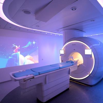
Philips now features videos with Disney characters as part of its Ambient Experience. Image courtesy of Philips Healthcare.Philips launched Ambient Experience in 2005 to give patients a more soothing experience during their imaging scans than a sterile radiology suite. The concept features lighting and music, with over 40 themes that can be selected by patients. The company recently marked the 2,000th installation of Ambient Experience, at a facility in Germany.
As part of the agreement, Philips will be coordinating a research study of the videos in use at six hospitals in Europe. Kids who undergo scans with the videos and their parents will fill out questionnaires asking them to describe their experience, and researchers will also track statistics like scan rescan rates, according to Werner Satter, general manager of healthcare experience solutions at Philips.
In other Ambient Experience news, Philips is highlighting results of a customer survey that found that 91% of Ambient Experience users are either likely or extremely likely to recommend the concept to other hospitals. In all, 76% of survey respondents reported that Ambient Experience decreased patient tension, 71% said it reduced fear, 66% said it induced patient calmness, and 63% said it improved patient cooperation during imaging exams.


![Overview of the study design. (A) The fully automated deep learning framework was developed to estimate body composition (BC) (defined as subcutaneous adipose tissue [SAT] in liters; visceral adipose tissue [VAT] in liters; skeletal muscle [SM] in liters; SM fat fraction [SMFF] as a percentage; and intramuscular adipose tissue [IMAT] in deciliters) from MRI. The fully automated framework comprised one model (model 1) to quantify different BC measures (SAT, VAT, SM, SMFF, and IMAT) as three-dimensional (3D) measures from whole-body MRI scans. The second model (model 2) was trained to identify standardized anatomic landmarks along the craniocaudal body axis (z coordinate field), which allowed for subdividing the whole-body measures into different subregions typically examined on clinical routine MRI scans (chest, abdomen, and pelvis). (B) BC was quantified from whole-body MRI in over 66,000 individuals from two large population-based cohort studies, the UK Biobank (UKB) (36,317 individuals) and the German National Cohort (NAKO) (30,291 individuals). Bar graphs show age distribution by sex and cohort. BMI = body mass index. (C) After the performance assessment of the fully automated framework, the change in BC measures, distributions, and profiles across age decades were investigated. Age-, sex-, and height-adjusted body composition reference curves were calculated and made publicly available in a web-based z-score calculator (https://circ-ml.github.io).](https://img.auntminnie.com/mindful/smg/workspaces/default/uploads/2026/05/body-comp.XgAjTfPj1W.jpg?auto=format%2Ccompress&fit=crop&h=100&q=70&w=100)


![Overview of the study design. (A) The fully automated deep learning framework was developed to estimate body composition (BC) (defined as subcutaneous adipose tissue [SAT] in liters; visceral adipose tissue [VAT] in liters; skeletal muscle [SM] in liters; SM fat fraction [SMFF] as a percentage; and intramuscular adipose tissue [IMAT] in deciliters) from MRI. The fully automated framework comprised one model (model 1) to quantify different BC measures (SAT, VAT, SM, SMFF, and IMAT) as three-dimensional (3D) measures from whole-body MRI scans. The second model (model 2) was trained to identify standardized anatomic landmarks along the craniocaudal body axis (z coordinate field), which allowed for subdividing the whole-body measures into different subregions typically examined on clinical routine MRI scans (chest, abdomen, and pelvis). (B) BC was quantified from whole-body MRI in over 66,000 individuals from two large population-based cohort studies, the UK Biobank (UKB) (36,317 individuals) and the German National Cohort (NAKO) (30,291 individuals). Bar graphs show age distribution by sex and cohort. BMI = body mass index. (C) After the performance assessment of the fully automated framework, the change in BC measures, distributions, and profiles across age decades were investigated. Age-, sex-, and height-adjusted body composition reference curves were calculated and made publicly available in a web-based z-score calculator (https://circ-ml.github.io).](https://img.auntminnie.com/mindful/smg/workspaces/default/uploads/2026/05/body-comp.XgAjTfPj1W.jpg?auto=format%2Ccompress&fit=crop&h=112&q=70&w=112)


