Abdominal imaging of COVID-19 patients has shown that bowel abnormalities, including ischemia, are common findings in these cases, according to research published online May 11 in Radiology. Cholestasis is also frequently present.
In a retrospective study, a team of researchers led by Dr. Rajesh Bhayana of Massachusetts General Hospital (MGH) reviewed abdominal imaging studies performed in 412 consecutive patients admitted to their institution and who had tested positive for SARS-CoV-2. They found that 17% of the patients had also received cross-sectional abdominal imaging, including 44 ultrasounds, 42 CT studies, and one MRI scan.
Of these patients, 31% of the CT scans -- representing 3.2% of all patients -- showed bowel abnormalities. These bowel abnormalities, which included thickening and findings of ischemia such as pneumatosis and portal venous gas, were more frequently experienced in intensive care unit (ICU) inpatients, according to the researchers. Surgical correlation in four patients revealed unusual discoloration of bowel in three of the patients and bowel infarction in two patients.
In two of the patients who had bowel resection, pathology results showed ischemia with patchy necrosis. Both of the patients had fibrin thrombi in submucosal arteries, suggesting that the bowel ischemia was caused by these thrombi, according to the researchers.
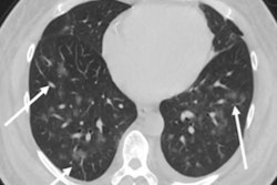
In addition, lung base findings resulted in a COVID-19 diagnosis in one patient who had presented with only abdominal symptoms. The researchers also found that 87% of right upper quadrant ultrasounds were performed due to liver laboratory findings, and 54% showed a dilated sludge-filled gallbladder suggestive of cholestasis.
"Some findings were typical of bowel ischemia, or dying bowel, and in those who had surgery we saw small vessel clots beside areas of dead bowel," Bhayana said in a statement from the RSNA. "Patients in the ICU can have bowel ischemia for other reasons, but we know COVID-19 can lead to clotting and small vessel injury, so bowel might also be affected by this."
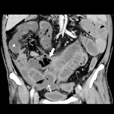
 Axial (A) and coronal (B) CT of the abdomen and pelvis with IV contrast in a 57-year-old man with a high clinical suspicion for bowel ischemia. There was generalized small-bowel distension and segmental thickening (arrows), with adjacent mesenteric congestion (thin arrow in B), and a small volume of ascites (* in B). Findings are nonspecific but suggestive of early ischemia or infection. Images courtesy of Radiology.
The spectrum of bowel findings in these COVID-19 patients could possibly be explained by direct viral infection, small vessel thrombosis, or nonocclusive mesenteric ischemia, according to the authors.
"ACE2 expression is most abundant in lung alveolar epithelial cells, enterocytes of the small intestine, and vascular endothelium suggesting that small bowel and vasculature may be susceptible to SARS-CoV-2 infection," they wrote.
Further study is required, however, to clarify the cause of bowel findings in COVID-19 patients and to determine whether SARS-CoV-2 plays a direct role in bowel or vascular injury, according to the researchers.