No signs of residual gadolinium uptake in the brain were found following intra-articular administration of a gadolinium-based contrast agent (GBCA) for MR arthrography in a new study, published online January 25 in Skeletal Radiology. The findings indicate that residual gadolinium may not occur with intra-articular use.
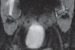
MR arthrography with gadolinium contrast is considered the gold standard for patients with intra-articular hip and shoulder issues. With the technique, diluted gadolinium -- at dose levels significantly lower than with intravenous use -- is injected within a joint to help distend the joint capsule and extend contrast into abnormalities.
However, recent studies have found increased signal intensity emanating from certain brain regions following the intravenous use of gadolinium with MRI scans. The question is, does this phenomenon also occur following intra-articular use?
To answer this question, a research team led by Katherine Bunnell and colleagues from Massachusetts General Hospital (MGH) retrospectively analyzed 109 consecutive patients (mean age, 44 ± 14 years) who had undergone at least one MR arthrogram of the shoulder (83 patients, 76%) or hip (26 patients, 24%) with the linear GBCA gadopentetate dimeglumine (Magnevist, Bayer HealthCare) between September 2014 and November 2017.
The mean dose of gadolinium was 0.08 (± 0.01) mL. Among the subjects, 100 patients (92%) underwent one scan, six patients (6%) underwent two MR arthrograms, and three patients (3%) had three arthrograms. None of these patients previously had been exposed to a GBCA.
The subjects later underwent 1.5-tesla or 3-tesla MRI brain scans (GE Healthcare or Siemens Healthineers), which included unenhanced T1-weighted sequences. The mean time between the initial MR arthrograms and the brain scans was 3.3 years (± 3.2 years). For comparative purposes, the researchers included 149 healthy control subjects (mean age, 43 ± 15 years), also with no past exposure to a GBCA.
To detect residual gadolinium, the researchers looked at four brain regions -- the pons, dentate nucleus, globus pallidus, and thalamus -- and cerebrospinal fluid (CSF). Signal intensities from the four brain regions were averaged and normalized to CSF to account for differences in magnet strengths and acquisition parameters and to calculate signal intensity ratios to estimate levels of gadolinium deposition.
In analyzing the results, the researchers found no statistically significant differences in mean signal intensity in any brain region in the MR arthrography group compared with control subjects.
| Signal intensity ratios of MR arthrography patients after GBCA use | |||
| Controls | MR arthrography patients | p-value* | |
| Pons/CSF | 2.03 ± 0.31 | 2.04 ± 0.29 | 0.7 |
| Dentate nucleus/CSF | 1.99 ± 0.32 | 2.02 ± 0.28 | 0.4 |
| Globus pallidus/CSF | 2.06 ± 0.30 | 2.06 ± 0.26 | 1.0 |
| Thalamus/CSF | 2.07 ± 0.30 | 2.10 ± 0.26 | 0.3 |
Multiple GBCA doses did not appear to influence mean signal intensity, as there were no statistically significant differences in signal intensity (p-value range, 0.2-0.5) between the nine patients (8%) who underwent more than one MR arthrogram and patients with only one contrast-enhanced MR arthrogram.
This statistical similarity between the number of GBCA doses and signal intensity ratios "suggests that intra-articular administration of GBCAs, using doses typically administered for [MR arthrograms], which are more than an order of magnitude smaller than typical doses of intravenous GBCAs, is not associated with intracranial gadolinium deposition," Bunnell and colleagues concluded.