Dear MRI Insider,
Evaluating large-vessel vasculitis (LVV) could soon become much more efficient. French researchers are reporting early success in combining MRI and PET to improve their analysis of abnormalities from the condition.
By merging contrast-enhanced anatomic assessments of MRI with the quantitative measurement of FDG uptake in PET, the researchers have accurately characterized and differentiated between inflammation and fibrosis in the aortic walls and auxiliary branches. This accumulation of data could help direct clinicians to more appropriate treatment for LVV and reduce the need for temporal artery biopsies. Get the details in our Insider Exclusive.
In other MRI news, Swiss researchers are using MRI to measure left ventricular hypertrophy to better predict death and heart failure related to coronary artery disease. Their findings show MRI can outperform coronary artery calcium scores from CT.
Gadolinium is in the news in Germany, where soft drinks served at fast-food outlets in a half-dozen cities were found to contain trace levels of the element. Researchers suspect the gadolinium comes from patients excreting gadolinium-based contrast after their MRI scans. Fortunately, the levels most likely do not represent a health risk.
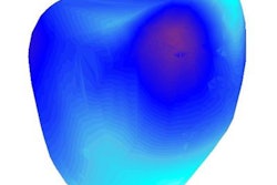
In the meantime, researchers from the U.K. have developed a new 3D MRI technique for calculating heart muscle strain without the need for gadolinium contrast. The image-registration algorithm uses computational mathematics to calculate heart muscle tracking and strain on 3D heart models based on contrast-free MRI scans.
Also, radiologists who used structured reporting to interpret MRI scans of glioma patients produced reports that were judged to be more consistent and easier to understand than conventional free-text reports. Structured reporting demonstrated superior traits across multiple domains, including greater consistency and completeness of history reporting, reduced ambiguity, and improved conciseness, researchers found.
Finally, virtual 3D models of the knee based on MRI scans of anterior cruciate ligament (ACL) tears revealed which regions in the knee were most likely to develop osteoarthritis. The technique could help individualize rehabilitation strategies for patients after ACL surgery.
Be sure to stay in touch with the MRI Community on a regular basis for news and research developments from around the globe.


