What caused a series of mysterious illnesses among diplomatic personnel at the U.S. embassy in Havana in 2016 and 2017? A study published February 15 in the Journal of the American Medical Association hints at symptoms similar to concussion but without evidence of trauma. And MRI scans were unable to shed light on the mystery.
News headlines in 2017 sounded like they were pulled from the Weekly World News: Personnel at the U.S. embassy in Havana were reported to be experiencing an array of neurological symptoms such as fatigue, vision problems, headache, and drowsiness. Many of them said the symptoms began after they reported hearing extremely loud, directional auditory and sensory stimuli that some described as sounding like "grinding metal" or "piercing squeals."
Embassy personnel had begun reporting the cases to their Havana medical unit in 2016, with more than 80 individuals undergoing evaluation between February and April 2017. Ultimately, some two dozen people were identified with exposure to what became known as "directional phenomena" and symptoms that resembled mild traumatic brain injury, also known as concussion. The case was referred to clinicians at the University of Pennsylvania's Center for Brain Injury and Repair; they reported their evaluation of 21 of the individuals in the new paper (JAMA, February 15, 2018).
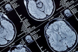
The embassy personnel received a battery of tests that included a full range of neuropsychological assessments, from auditory attention to mood functioning and effort. MRI scans were performed on a 3-tesla scanner on all 21 individuals (Magnetom Prisma, Siemens Healthineers). Four patients who reported unilateral peripheral vestibulopathy (vertigo) also received MRI scans with a focus on the auditory canals.
Eighteen of the 21 individuals had reported hearing a "novel, localized sound" in their homes or hotel rooms, with symptoms beginning immediately thereafter in 20 cases. They described the sounds as "directional, intensely loud, and with pure and sustained tonality." Some also reported pressure or vibratory stimuli, similar to "air baffling" in a car with the windows rolled down, wrote lead author Dr. Randel Swanson, PhD, and colleagues.
In addition to symptoms such as fatigue and irritability, the clinical evaluations raised concerns about cognitive impairment in 16 individuals. In the six people with complete neuropsychological testing and data analysis, all had "significant areas of cognitive weakness and/or impairment," the group noted. Two individuals met the criteria for post-traumatic stress disorder.
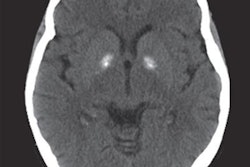
Unfortunately, the MRI scans did little to illustrate what was afflicting the personnel. Most of the 21 patients had "conventional imaging findings," with nine demonstrating a few small, nonspecific foci in the white matter that were bright on T2 imaging. Three patients had multiple white-matter foci on T2 scanning, but the findings were not specific to the directional phenomena they experienced and could have been the result of pre-existing conditions or other disease processes, according to the group. MRI scans of the four patients with unilateral peripheral vestibulopathy were normal.
The researchers concluded that they were baffled as to whether the clinical symptoms were related to the directional phenomena the individuals had reported. Although the symptoms were closest to those experienced by individuals with concussion, there were major differences, such as ear pain, tinnitus, and vertigo. Swanson and colleagues even went so far as to suggest that there could be some kind of new pathology at work.
"The clinical manifestations may represent a novel clinical entity, which appears to have resulted from a widespread brain network dysfunction ... as seen in mild traumatic brain injury, or concussion," they wrote. "It is currently unclear if or how the noise is related to the reported symptoms."