Monday, December 1 | 3:50 p.m.-4:00 p.m. | SSE14-06 | Room E450B
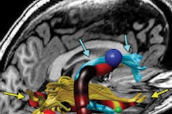
Whole-body MR neurography (MRN) is a feasible way to evaluate the disease burden of neuropathy, or damage to the peripheral nervous system, according to research to be presented in this afternoon session.The study was led by Dr. Avneesh Chhabra, an associate professor of radiology and orthopedic surgery at UT Southwestern Medical Center at Dallas.
MRN benefits patients because it is a noninvasive way to assess the effects of the disease, which include nerve thickening, in a one-hour scan, Chhabra and colleagues concluded.
The study included 11 patients with neuropathy and 18 control subjects who were imaged on a 3-tesla MRI scanner to examine differences among the different types of diffuse neuropathies.
The researchers acquired two or three sets of 3D MRN images from the base of the skull to the proximal thigh, as well as contiguous axial T2-weighted spectral-attenuated inversion recovery (SPAIR) imaging of the symptomatic extremity and diffusion-tensor imaging (DTI) of the brachial and lumbosacral plexuses.
Two readers assessed the quality of the images and obtained measurements, including nerve diameter and signal intensity ratios for various nerves.
MRN showed that nerve thickening was significant in the lumbosacral plexus, sciatic, and femoral nerves. There was also significant hyperintensity in brachial and lumbosacral plexuses. Nerve entrapments in the extremities were seen in four of the 11 subjects.
Whole-body MRN is a "feasible method with good to excellent interobserver performance that can be objectively used to evaluate disease burden and detect differences among diffuse neuropathies," the group concluded.