Monday, December 1 | 11:40 a.m.-11:50 a.m. | SSC10-08 | Room N226
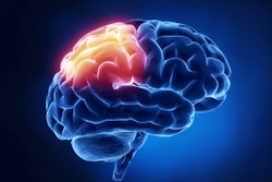
Researchers at University Hospital Essen in Germany are using the power of 7-tesla MRI to gather more information on patients with glioblastoma multiforme.The goal of their study was to compare the diagnostic ability of 3- and 7-tesla MRI for patients with this most common and aggressive malignant primary brain tumor.
The group evaluated10 patients using a 3-tesla MRI system (Magnetom Skyra, Siemens Healthcare) and a 7-tesla whole-body MRI system (Magnetom 7T, Siemens) with 32-channel head coils.
Field strength comparisons were performed for a variety of MRI sequences, including susceptibility-weighted imaging, T2-weighted fluid-attenuated inversion recovery (FLAIR), and postcontrast T1-weighted 3D magnetization-prepared rapid acquisition with gradient echo (MPRAGE).
Two radiologists assessed tumor microvasculature, potential necrosis, and edema, as well as overall image quality for all sequences.
The readers found equally high delineation of tumor extent, morphology, and edema at both magnet strengths. However, 7-tesla SWI was significantly better than 3-tesla MRI for assessing tumor-associated microvasculature.
"The combination of increased signal-to-noise ratio, enabling higher spatiotemporal resolution imaging, and susceptibility effects at 7 tesla have been shown to enable improved assessment of tumor delineation and tumor-associated vasculature," presenter Dr. Lale Umutlu told AuntMinnie.com.
The study is ongoing, and the researchers hope to include additional tumors in the near future, he added.