Monday, December 1 | 11:30 a.m.-11:40 a.m. | SSC09-07 | Room N229
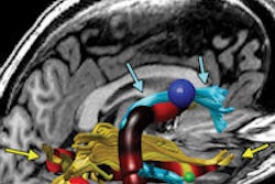
Researchers from Massachusetts General Hospital (MGH) and Harvard Medical School have developed an MRI technique that could assist with prognoses for patients who are comatose due to traumatic brain injury.The method combines diffusion-tensor MRI (DTI-MRI) with a Tracts Constrained by Underlying Anatomy (TRACULA) technique, which automatically reconstructs white-matter pathways from diffusion-tensor images.
"This research presents a method to quantify an injury to white-matter tracts in traumatic brain injury," explained lead author Dr. Emad Ahmadi, a research fellow in MGH's department of radiology. "The goal is to use this method to predict the prognosis of coma caused by severe traumatic brain injury."
In the study, Ahmadi and colleagues used the imaging method to quantify and localize 18 white-matter bundles in 53 patients who were comatose for at least seven days to see if they could predict neurologic and cognitive outcomes. The study included 17 control subjects for additional comparisons.
Thirteen white-matter bundles showed significant differences in at least in one region of the brain between the comatose patients and control subjects, according to the researchers. In addition, 11 white-matter bundles showed significant difference in at least one region between patients with good and poor outcomes.
"We have shown that the extent of white-matter injury is underdiagnosed by conventional MRI sequences," Ahmadi explained. "Other imaging modalities such as CT don't actually give that much information about white-matter injury."
The group is continuing the research by studying the injuries to individual white-matter tracts and their effects on neurological and psychological deficits.
"The ultimate goal is to predict specific neuropsychological deficits that the patients will have using an imaging protocol," he said. "Ideally, this imaging protocol will be acquired in the acute phase after traumatic brain injury, will determine white-matter injuries, and will predict what neuropsychological deficits the patient will experience."
That information could help clinicians make therapeutic decisions such as whether or not to continue intensive care for comatose patients, Ahmadi said.