In a comparison of 3-tesla MR imaging techniques of the spine, German researchers have found that a technique using a parallel radiofrequency (RF) transmission technology produces diagnostic-quality images equal to conventional MR images in approximately one-third the time.
The study from the University of Bonn showed that among its patient sample, scan time for a whole-spine examination could be reduced by 31% through the parallel imaging technique. The research was presented at the 2010 annual RSNA meeting.
Parallel transmission, also known as multitransmit, uses specialized RF coils in which each element of a phased-array coil is associated with a dedicated RF channel, which can be processed and combined for faster MR imaging. This enables the frequency, amplitude, phase, and waveform to be automatically adjusted for optimal uniformity for each patient's anatomy.
The technique can speed up imaging time and also reduce artifacts, so the researchers decided to apply it to spine imaging, according to lead study author Michael Nelles, MD, from the university's department of radiology.
"The spine can be quite challenging in MR imaging," Nelles said. "So, we were curious about the advantages that may arise from the use of parallel transmission at 3-tesla MRI of the spine."
In the prospective study, researchers scanned 30 consecutive patients between November 2008 and January 2009 with and without the use of parallel transmission. The patient sample included 19 women and 11 men, with a mean age of 50 years (range, 25 to 80 years).
MRI protocol
The researchers used a 3-tesla MRI system (Achieva 3.0T TX, Philips Healthcare, Andover, MA) and RF amplifiers capable of operating in both conventional single-transmission and parallel-transmission modes. A total of 77 MR image sequences with and without parallel transmission were obtained for comparison.
"To perform a qualitative analysis, two independent readers compared the diagnostic image quality of the single and multitransmit modes according to a simple four-rating scheme," said Nelles. In addition, image-to-contrast ratios were calculated between reference tissues and vertebrae to evaluate the single- and dual-source transmission results.
The researchers' analysis concluded that total scan time for a whole-spine examination could be reduced by 31% with the use of dual-source transmission. The average time for parallel scanning was 30 minutes and 16 seconds, compared with 44 minutes in single-source mode.
Reduced scan time
"If you add up the scan times for the individual sagittal scans," Nelles added, "it will result in over half an hour of acquisition time for the single transmit mode and approximately 23 minutes for the parallel transmit mode."
Spinal 3-tesla MRI with parallel transmission also yielded a median assessment of at least adequate image quality in all of the compared sequences. There were no cases in which an image was determined to be of nondiagnostic quality.
 |
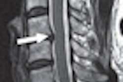
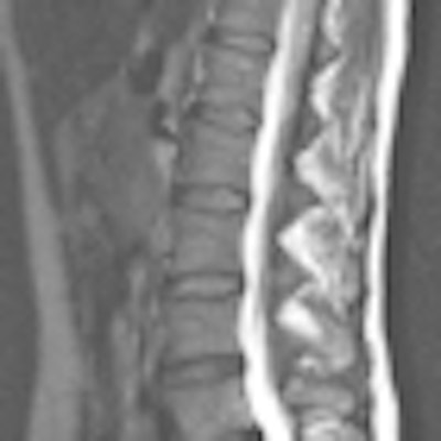
| The lumbar spine of a 68-year-old woman with sequestered disk herniation is shown in parallel transmission mode (right) and single-source transmission mode (left). Independent readers rated the parallel image as superior. Images courtesy of Radiology (December 2010, Vol. 257:3, pp. 743-753). |
With the "significant acceleration" of scan time and parallel transmission with 3-tesla MRI of the spine, Nelles said, there is "really a strong advantage for patients with back pain and in cases of claustrophobia. The diagnostic image quality of these fast sequences stays comparable to that of the single standard transmission sequences."
Nelles added that he doubted the parallel transmission technique could be used to see bone metastasis or lesions along the spine that were not identifiable through the single-source mode, although he and his colleagues had not researched this issue.
By Wayne Forrest
AuntMinnie.com staff writer
January 11, 2011
Related Reading
Multitransmit MR improves lumbar spine imaging at 3 tesla, December 9, 2010
Stand-up MRI detects impact of heavy backpacks on children, January 6, 2010
MRI reveals high incidence of disk disease in overweight and obese youngsters, December 2, 2009
MRI finds proximal femur fractures missed by x-ray, September 1, 2009
MRI helps find fractures among elderly female ED patients, April 29, 2009
Copyright © 2011 AuntMinnie.com