Postmenopausal women, including those older than 70 years of age, who have been newly diagnosed with cancer in one breast have higher cancer detection rates when the other breast is scanned for tumors with MRI compared to premenopausal women, according to a new study in the Breast Journal.
Lead author Dr. Johnny Ray Bernard Jr., a radiation oncologist at Mayo Clinic in Jacksonville, FL, and colleagues also found that women 70 years of age and older had a higher prevalence of cancer in the second breast as detected by MRI than younger patients. MRI detected cancer in the second breast in seven (5.4%) of the 129 elderly women in the study (TBJ, March/April 2010, Vol. 16:2, pp. 118-126).
"This study was specific in that it didn't just look at the opposite breast," Bernard said. "We also scanned the same breast, because we think MRI can detect additional disease in the same breast that may be more extensive than what is seen on the mammogram. That helps a surgeon with planning for the initial breast where the cancer was diagnosed."
Patient sample
Mayo Clinic researchers retrospectively reviewed records of 425 women who underwent bilateral breast MRI at the Jacksonville facility between February 2003 and November 2007. Of the 425 women, 129 (30%) of them were 70 years of age or older at the time of their diagnosis. The overall median age for all 452 women was 62 years, ranging from 25 to 91 years.
All women underwent bilateral contrast-enhanced breast MRI within 60 days of a new, histologically confirmed diagnosis of breast cancer. Contrast-enhanced MRI was performed using a 1.5-tesla scanner (Espree, Symphony, or Avanto, Siemens Healthcare, Malvern, PA) and a dedicated breast surface coil. Sagittal and axial 3D T1-weighted high-resolution images were obtained before and after contrast administration, including initial and delayed enhancement sequences.
Postmenopausal status
In the review of patient records, researchers found that postmenopausal status was the only statistically significant predictor of contralateral cancer detected by MRI. A contralateral biopsy was recommended and performed on the basis of MRI alone in 72 of 425 women (17%), with contralateral carcinoma confirmed pathologically in 16 of the 72 women (22%) for an overall diagnostic yield of 3.8%.
Researchers also found that invasive carcinoma was detected in 13 women and ductal carcinoma in situ (DCIS) was found in three women. MRI was able to detect contralateral carcinomas ranging in size from 0.3 cm to 3.0 cm, with a median size of 0.8 cm.
In the group of women older than 70 years of age, biopsy was recommended and performed based on the MRI results alone in 16 of 129 women (12%). Of these 16 women, seven (44%) had pathologically confirmed carcinoma. Five of the seven women were diagnosed with invasive carcinoma, while two women were found to have DCIS.
|
Mayo Clinic in Jacksonville traditionally has used MRI as an adjunct to mammography. "Particularly in women with dense breasts, mammograms have a difficult time trying to discern between breast tissue and what is abnormal," Bernard said. "With MRI, the picture comes out more clearly, so it is usually easier for MRI to distinguish between dense tissue and abnormalities."
One of the criticisms of MRI, however, is that it may subject women to unnecessary biopsies, because the modality is so sensitive and may pick up lesions that are not cancer.
"If you think that false positives are more damaging than finding cancers, then you will say MRI is not needed. If you think finding these cancers is more important than the false positives from MRI, then you will say MRI is a great exam," he added. "For us at Mayo, we believe the benefit of finding these cancers is worth putting these women through the MRI, though we know some of them may have biopsies that will not come back with cancer."
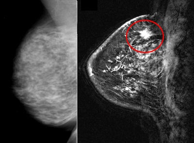 |
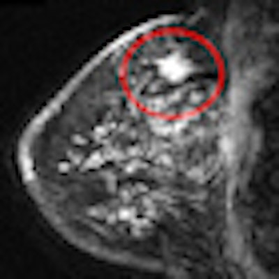
| A 76-year-old woman with 1.4-cm contralateral lesion noted on MR (right) after a negative mammogram (left). Image courtesy of Dr. Johnny Ray Bernard Jr. |
Bernard and his colleagues noted that even elderly women in good health can potentially benefit from earlier detection of breast cancer, and MRI screening of the undiagnosed breast should be considered in all postmenopausal women diagnosed with a breast cancer.
The study findings could help reduce healthcare costs by limiting screening MRI to patient groups likely to have greater detection rates, such as the postmenopausal women in this study, and by having both cancers treated at the same time instead of undergoing potentially toxic and expensive treatments more than once, he said.
By Wayne Forrest
AuntMinnie.com staff writer
March 17, 2010
Related Reading
Breast MRI CAD yields high sensitivity, specificity, March 8, 2010
Radiation exposure increases risk of breast cancer, March 4, 2010
Breast MRI excels at high-risk screening; mammo not needed, February 26, 2010
Breast MRI best for finding mammographically occult cancers, November 9, 2009
Breast MRI can resolve mammography findings, but use with caution, October 6, 2009
Copyright © 2010 AuntMinnie.com


