Australian investigators are heading up a research project that will look at the viability of MR spectroscopy analysis of primary breast lesions. They presented their concept last week in Orlando at the Era of Hope meeting, sponsored by the U.S. Department of Defense Breast Cancer Research Program.
This pre-surgical methodology will separate benign tumors from malignant ones, and determine if cancer has spread to the lymph nodes based on the altered cellular chemistry of tumors. The plan is to combine MRS with a statistical classification strategy (SCS) to analyze tissue aspirated from fine-needle biopsy. Imaging of the specimen will be performed with a custom-made bench-top 8.5-tesla MR unit designed by Magnex Scientific of Yarnton, U.K.
The software and hardware for the analysis is being developed by Spectex of Sydney and Bruker Biospin of Karlsruhe, Germany. The software includes automated data collection, quality assurance parameters, and SCS-base classifiers.
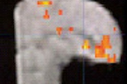
As with other spectroscopy studies of the breast, the presence or absence of two chemicals, choline and creatine, will be studied. Both are chemical indicators of malignancy: creatine is always found in malignant tissue, while high levels of choline are generally indicative of malignancy. But the combination of MRS and the classification strategy will go a step further than previous MRS studies.
"This looks at all of the chemical information rather than just the choline and creatine. The study that we’ve presented here is based on a number of chemical regions in the spectrum," explained Cynthia Lean, Ph.D., in an interview with AuntMinnie.com. Lean is the scientific director of the Institute for Magnetic Resonance Research in Sydney.
The study will build on previous research done by Lean and her colleagues. In a 1997 article published in Radiology, they reported the results of 218 fine-needle biopsy specimens that were analyzed with MRS. In this case, they were only testing to see if MRS could differentiate between benign and malignant cancer.
They found that invasive carcinoma produced an increased signal at 3.25 ppm that could be attributed to choline-containing metabolites. They came up with a sensitivity of 95% and a specificity of 96% for this technique (Radiology, September 1997, Vol. 204:3, pp. 661-666).
Lean said her group expects the more extensive study to produce the same results with regard to the distinction between benign and malignant tumors. "However, using the stastistical classification strategy to extract that additional information allows us to predict whether or not lymph nodes are involved from the cells from the primary tumor. And that’s the important thing," she said.
Testing is scheduled to begin in 2003 at two independent sites: Beth Israel Deaconess Medical Center in Boston and the Karolinska Institute in Stockholm. Acceptance testing of the Spectex system will be done at Lean’s institute. The patient population will be determined by the clinicians at each institution. The criterion for inclusion is a palpable lesion, or one that can be identified with ultrasound so that pathological information can be obtained, Lean said.
Lean said this MRS method could serve as an adjunct to sentinel node biopsy. MRS support for the veracity of a negative sentinel node biopsy could reduce the need for axillary surgery and possible lymphodaema.
By Shalmali PalAuntMinnie.com staff writer
October 1, 2002
Related Reading
MRI useful in women with early-stage breast cancer, September 30, 2002
MRS tops MRI in staging hypoxic injury, August 16, 2002
Prototype MR elastography reveals mechanical properties of breast cancer, July 25, 2002
Smaller fields of view cut MR spectroscopy scan time, December 20, 2001
Copyright © 2002 AuntMinnie.com