A new study published in EJNMMI Research found that SPECT/CT imaging using Tc-99m MIP-1404 is a practical and cost-effective alternative to PET/CT for staging high-risk prostate cancer, achieving 91% agreement on treatment decisions while offering broader accessibility and significantly lower costs.
- SPECT/CT Performance: Achieved 81% sensitivity for lymph node metastasis and 85% for bone metastases with 100% specificity compared to PET/CT
- Treatment Decision Alignment: Therapy decisions based on SPECT/CT agreed with PET/CT in 91% of the 33 patients studied
- Cost and Accessibility: SPECT/CT costs three to four times less than PET/CT and requires no cyclotron or generator dependency
- Clinical Concordance: Classification of metastasis burden was concordant in 85% of cases with SPECT/CT versus 64% with conventional imaging
- Study Design: Phase II prospective study comparing Tc-99m MIP-1404 CZT SPECT/CT versus Ga-68 PSMA-11 PET/CT in newly diagnosed high-risk prostate cancer patients
SPECT/CT may represent a practical alternative to PET/CT for primary staging in patients with high-risk prostate cancer, according to a study published July 22 in EJNMMI Research.
The finding is from a phase II prospective study that compared technetium-99m (Tc-99m)-MIP-1404 CZT SPECT/CT versus gallium-68 (Ga-68) prostate-specific membrane antigen (PSMA)-11 PET/CT among 33 patients with newly diagnosed disease, noted lead author Aws Saudi, MD, of Linköping University in Sweden, and colleagues.
“Considering its broader accessibility, Tc-99m MIP-1404 CZT SPECT/CT may represent a practical alternative for primary staging, especially in settings with limited PET/CT availability,” the group wrote.
PSMA PET/CT has demonstrated superior accuracy for primary prostate cancer staging compared with conventional imaging, but access is constrained by cyclotron requirements, generator dependency, and costs up to three to four times higher than SPECT/CT, the authors explained. The development of Tc-99m-based PSMA-targeted agents may offer a potential route to PSMA-quality staging in settings where PET/CT infrastructure is limited, they hypothesized.
To test the technique, the researchers prospectively enrolled 33 men aged over 50 years old with newly diagnosed high-risk prostate cancer between January 2023 and December 2024. All participants underwent three scans within a 30-day interval: Ga-68-PSMA-11 PET/contrast-enhanced (CE) CT, Tc-99m MIP-1404 CZT SPECT/CT, and Tc-99m HDP CZT SPECT/CT (bone scan). Images were analyzed using the prostate-specific membrane antigen reporting and data system (PSMA-RADS) version 2.0.
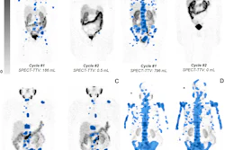
 Maximum intensity projection of Ga-68 PSMA-11 PET/CE-CT (A), Tc-99m MIP-1404 CZT SPECT/CT (B) and Tc-99m HDP CZT SPECT/CT (C) imaging. Ga-68 PSMA-11 PET/CE-CT and Tc-99m MIP-1404 CZT SPECT/CT showing tracer uptake in lymph nodes below the iliac bifurcation and above, paraaortic and left supraclavicular, bone uptake in the right proximal femur and right scapula (arrows). Tc-99m HDP CZT SPECT/CT did not show any uptake suspicious for metastatic disease. No sclerotic lesions suspicious of metastases were detected by the CT component in any of the studies. Fusion axial imaging of Ga-68 PSMA-11 PET/CE-CT (D) and Tc-99m MIP-1404 CZT SPECT/CT (E) showing uptake in lymph nodes in the left pelvic area below the iliac bifurcation (arrows). Fusion axial imaging of Ga-68 PSMA-11 PET/CE-CT (F) and Tc-99m MIP-1404 CZT SPECT/CT (G) showing uptake in the right proximal femur, not detected by Tc-99m HDP CZT SPECT/CT (H), (arrows). EJNMMI Research
At the lesion level, Ga-68-PSMA-11 PET/CE-CT detected significantly more lymph node metastases than Tc-99m MIP-1404 SPECT/CT (mean 8.19 versus 2.09 per patient; p < 0.001), while no significant difference was found for skeletal lesion counts (p = 0.318). Further, classification of metastasis burden into high volume metastasis and low volume metastasis was concordant in 85% of cases based on Tc-99m MIP-1404 CZT SPECT/CT compared to Ga-68 PSMA-11 PET/CE-CT, while conventional imaging agreed in 64% of cases.
Lastly, therapy decisions based on Tc-99m MIP-1404 CZT SPECT/CT were concordant with those based on Ga-68 PSMA-11 PET/CE-CT in 91% (30/33) of patients.
“It is important to highlight that despite the higher lesion count with Ga-68 PSMA-11 PET/CT, this did not translate into a meaningful change in treatment decisions in our study cohort,” the group wrote.
Further studies with larger cohorts are needed to strengthen its diagnostic performance and confirm the potential of Tc-99m MIP-1404 CZT SPECT/CT, the researchers concluded.
Read the full study here.