NEW ORLEANS – F-18 FDG-PET/MRI can identify previously unknown foci that generate chronic spinal pain and enable targeted interventions, according to a June 22 presentation at the Society of Nuclear Medicine and Molecular Imaging 2025 annual meeting.
Fatemeh Rashidi, MD, a postdoctoral research associate at the University of Wisconsin-Madison, discussed a study involving 23 patients with chronic spinal pain refractory to several standard-of-care therapies who underwent simultaneous PET/MRI scans.
“PET/MRI changed management in patients who had chronic spinal pain on average five years and were referred from CT or MRI and many surgeries, but no pain reduction after the procedures,” she said.
Chronic spinal pain, low back, and neck pain, cause significant disability and cost the U.S. healthcare system $134.5 billion annually, Rashidi explained. CT and MRI, which rely on morphologic abnormalities to identify the sources of pain, suffer from poor diagnostic accuracy and often lead to undesirable and harmful outcomes, such as unnecessary or ineffective surgeries and systemic treatments with morbid side effects, such as opioids, she added.
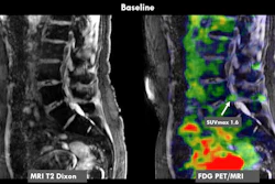
Conversely, F-18 FDG-PET has been shown to accurately identify pain generating foci by identifying the inflammation linked to painful conditions. Given that MRI provides high-resolution images with excellent soft tissue contrast that complement functional information on PET, in this study, Rashidi and colleagues evaluated the impact of the technique on managing patients with chronic spinal pain.
Imaging exams were reviewed in consensus by a dual-board-certified radiologist/nuclear medicine physician and a musculoskeletal radiologist, where abnormal F-18 FDG uptake on PET (uptake greater than expected for normal physiology) was correlated with MRI, and suspected foci of pain generation were identified. The group determined patients’ baseline pain on the Numeric Pain Rating Scale (0 = no pain, 10 = worst pain possible), as well as treatment decisions based on the PET/MRI scans and subsequent follow-up pain scores.
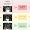
 Coronal fused FDG-PET/MRI image of a patient with right upper back pain demonstrates asymetric uptake in the right trapezius muscle without abnormality on MRI to suggest edema or denervation injury. Trigger point lidocaine injection of the area based on PET/MRI findings resulted in complete relief of symptoms (pain score reduced to 0 from a baseline of 6).Fatemeh Rashidi, MD, and SNMMI.
Out of the 23 patients, the pain region was the neck for five, upper back for four, and lower back for 14. PET/MRI changed management in 19 out of 23 patients, Rashidi reported. Eighteen underwent anesthetic/steroid injections, and one patient underwent surgery. The average follow-up pain score was significantly lower after the interventions (2.8) compared to baseline (5.6), and 13 patients reported 30% or greater reduction of pain.
“Given the high prevalence of chronic spinal pain, if even a fraction of patients receive an accurate diagnosis, FDG-PET/MRI's impact would be significant,” Rashidi noted.
Limitations of the study included the small sample size and lack of a control group. Thus, the true impact of F-18 FDG-PET/MRI on clinical management of chronic pain patients needs to be verified in larger, multi-center studies with appropriate controls, Rashidi concluded.