Replacing F-18 sodium fluoride (NaF) PET/CT imaging with F-18 prostate-specific membrane antigen (PSMA) PET/CT can improve treatment decisions among men with newly diagnosed prostate cancer, researchers in Denmark have reported.
Among 160 patients with high-risk disease, the switch resulted in changes to treatment plans for 13%, noted lead author Claus Madsen, PhD, of Copenhagen University, and colleagues.
“The use of F-18 PSMA-PET/CT resulted in significant metastatic stage migration and influenced treatment planning in a substantial proportion of patients,” the group wrote. The study was published October 9 in the Journal of Nuclear Medicine.
Diagnostic imaging of soft tissue such as lymph nodes and bone in patients with newly diagnosed prostate cancer is crucial for treatment planning, as the disease is highly metastatic to these sites. While F-18 PSMA-PET/CT has demonstrated superior accuracy in this setting compared with conventional imaging techniques, the impact of replacing F-18 NaF-PET/CT with F-18 PSMA-PET/CT remains underexplored, the authors explained.
To bridge the knowledge gap, the researchers analyzed imaging results from 160 patients with newly diagnosed high-risk disease who underwent both F-18 NaF-PET/CT and F-18 PSMA-PET/CT within three weeks. The F-18 PSMA-PET/CT results were initially withheld, with staging and treatment decisions based solely on F-18 NaF-PET/CT.
At later time points during multidisciplinary team conferences involving urologists, an oncologist, a nuclear medicine specialist, a radiologist, and a pathologist, staging and treatment plans were reassessed based on the F-18 PSMA-PET/CT results.
In total, 40 patients (25%) experienced a change in metastatic stage after F-18 PSMA-PET/CT results were added to the evaluation, compared with staging based on F-18 NaF-PET and CT results alone, the researchers reported. Thirty-eight patients (24%) were classified as having a more advanced stage, and two patients (1%) were classified as having a less advanced stage (p < 0.001).
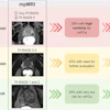
 Primary staging of a patient with high-risk prostate cancer. Initial F-18 NaF-PET/CT showed no regional or distant metastases. The patient later underwent radical prostatectomy with extended pelvic lymph-node dissection. In a blinded project review, F-18 PSMA-PET/CT revealed multiple intrapelvic lymph node metastases (blue arrows) and 1 lymph node metastasis near bifurcation of right common iliac artery (red arrows). Primary prostate lesion is also visible (green arrow). Three months postoperatively, the patient’s PSA remained elevated (0.67 ng/mL), and bicalutamide therapy was started.Journal of Nuclear Medicine
“These findings illustrate the potential consequences of implementing a shift in daily clinical practice from conventional imaging modalities (such as F-18 NaF-PET or bone scintigraphy and CT or MRI) to the more sensitive F-18 PSMA-PET/CT,” the researchers wrote.
Ultimately, the use of PSMA-PET/CT in clinical practice represents a paradigm shift in the staging of prostate cancer, with groups such as the European Association of Urology and the Society of Nuclear Medicine and Molecular Imaging recommending its use for primary staging in men with advanced disease, the group noted.
Nonetheless, to their knowledge, this was the first prospective study to assess stage migration and the tentative clinical consequences of replacing F-18 NaF-PET/CT with F-18 PSMA-PET/CT, they added.
“These findings are important, as PSMA-PET/CT becomes increasingly integrated into international guidelines for primary staging,” the researchers concluded.
The full study is available here.