PET/MRI and dual-energy CT (DECT) have revealed heart and lung abnormalities in patients with long COVID, according to a study published April 30 in the Journal of Nuclear Medicine.
The findings are from patients with cardiopulmonary symptoms lasting on average 303 days after infection, noted lead author Maria Trivieri, MD, PhD, of the Icahn School of Medicine at Mount Sinai in New York City, and colleagues.
“We posit that these findings warrant closer monitoring to exclude long-term sequelae, such as valvular disease or pulmonary hypertension,” the group wrote.
The COVID-19 pandemic resulted in more than 80 million infections and approximately 1 million deaths in the U.S. Beyond the acute phase of infection, long COVID has emerged as a distinct syndrome, manifesting in up to 69% of cases, the authors explained. The syndrome is characterized by symptoms lasting for four or more weeks after initial severe infections.
To further investigate the effects of long COVID, the group conducted the first multimodality, imaging-based assessment of the syndrome, with the largest cohort and longest interval from infection to imaging to date, they noted.
The researchers recruited a prospective cohort of 98 adults with a self-reported history of COVID-19 infections who were referred from outpatient clinics between December 2020 and July 2021. The most common symptom was shortness of breath (80%), and 27% of participants were hospitalized.
Participants underwent cardiopulmonary F-18 FDG PET/MRI and DECT imaging of the lungs. For comparison, the group also imaged a control group that included subjects with severe acute COVID-19 infection but without cardiopulmonary symptoms.
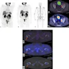
 Myocardial involvement. Representative examples show fused PET/MRI images in patients with findings suggestive of myocarditis. (A and B) Short-axis view of increased F-18 FDG uptake in basal septum segments (A), colocalized with area of increased T2 values (B) on parametric mapping. (C and D) Prominent F-18 FDG uptake in middle anterolateral and inferolateral walls (C), matching area of increased T1 values (D). (E and F) Short-axis views of avid F-18 FDG uptake in basal inferior and inferolateral walls (E), matching areas of increased extracellular volume (F). SUV scale is 0-5 g/mL. Journal of Nuclear Medicine
According to the results, PET/MRI scans were abnormal for 52 (57%) of the subjects: 22 showed cardiac involvement suggestive of myocarditis, 20 presented uptake reminiscent of pericarditis, 10 showed periannular uptake, and 28 showed vascular uptake (aortic or pulmonary). DECT revealed abnormalities in 63 (90%) of the subjects, with 47 demonstrating pulmonary infiltrates and 41 abnormal perfusion.
There was no myocardial, pericardial, periannular, or pulmonary uptake on the PET/MRI scans of the control group.
“In [long-COVID] subjects evaluated up to a year after coronavirus disease 2019 infection, our results indicate a high prevalence of abnormalities on PET/MRI and DECT,” the researchers wrote.
In addition, over a median follow-up period of 3.8 years, 15 (15%) of the participants experience cardiovascular events, such as heart failure, mitral valve disease, myocardial infarction, and pulmonary embolism, with 10 (67%) of these subjects having abnormal PET/MRI scans, the researchers noted.
Ultimately, because most patients had no prior scans or testing available for comparison, the researchers could not confidently rule out that some of the imaging abnormalities were preexisting. However, to minimize this, they noted that they excluded patients with any active disease before infection or with a temporal correlation between vaccines and symptoms.
“Further studies with larger cohorts, including larger control groups, are required to establish the generalizability of our findings,” the researchers concluded.
The full study is available here.