PET/MRI is among the newest technologies in radiology, and on its cutting edge is research on breast cancer imaging, according to a presentation at the International Society for Magnetic Resonance in Medicine (ISMRM) annual meeting.
In a June 6 session, Jonathan McConathy, MD, PhD, of the University of Alabama at Birmingham described studies at his institution using PET/MRI in women diagnosed with human epidermal growth factor receptor 2-positive (HER2+) breast cancer, among the most aggressive types.
"There's a lot of research in PET/MR, and areas we're interested in include breast cancer," McConathy said.
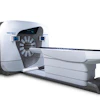
Both PET and MRI scanners are used in cancer imaging, with PET/MRI hybrid scanners developed about 12 years ago. These scanners can provide almost simultaneous detailed tumor assessments from MRI with complementary physiologic information from PET. Thus, the approach provides diagnostic value for clinicians not possible with either approach alone.
At McConathy's hospital, a multidisciplinary team is exploring the use of the technology to visualize whether women with HER2+ tumors are responding to treatment with trastuzumab (Herceptin), a monoclonal antibody specifically developed to target cancer in these patients. The drug was approved in the U.S. in 1998.
The nontherapeutic component of trastuzumab can be labeled with a zirconium-89 (Zr-89) radioisotope (chemically synthesized in laboratories) so that when the resulting PET radiotracer is injected into patients, it reveals activity at sites where its therapeutic counterpart may or may not be killing tumor cells. This allows clinicians to detect a patient's response to therapy.
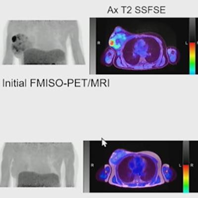
 PET/MRI hypoxia imaging in a patient with breast cancer. Image courtesy of Dr. Jonathan McConathy.
McConathy presented a case of a patient whose cancer was originally suspected to be HER2+ but who had a Zr-89 PET/MRI scan to confirm the diagnosis. The imaging revealed only a few lymph nodes showing signs of the disease in the supraclavicular lymph nodes (above the clavicle), and so HER2+ cancer was ruled out.
"That suggests she might not be a good candidate for further HER2+-targeted therapy," he said.
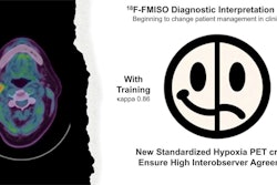
In addition, McConathy and colleagues are also exploring the use of PET/MRI with the radiotracer F-18 fluoromisonidazole (FMISO) for hypoxia imaging in women with breast cancer. Hypoxia in these cases refers to reduced oxygen in tissue around tumor sites, with the tracer designed to bind to these areas.
In one case McConathy presented, PET/MRI showed images of a patient with a large tumor, with FMISO-PET showing hypoxia at the molecular level, while MRI shows a large amount of "diffusion restriction" preventing the movement of water molecules in tissue at the site. In addition, a follow-up PET/MRI showed the patient's response to treatment.
"The patient actually responded quite well to chemotherapy ... this is kind of a dramatic image," he noted.
Ultimately, these are early days for PET/MRI in breast cancer, with applications of the technology in other cancers much more advanced, particularly in patients with prostate cancer, McConathy noted.
Nonetheless, while the advantages of PET/MRI are clear, limits remain.
"Complexity, cost, and reimbursement continue to be barriers to widespread PET/MRI for body oncology," he concluded.